Products
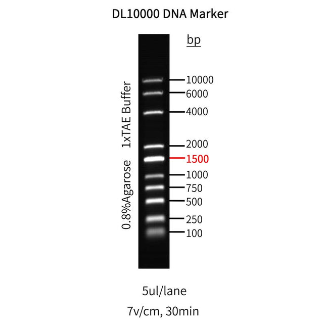
10000bp DNA Marker, 100-10000bp
Product description This product contains a DNA molecular weight marker consisting of 10 linear double-stranded DNA fragments at the following sizes: 100 bp; 250 bp; 500 bp; 750 bp; 1,000 bp; 1,500 bp; 2,000 bp; 4,000 bp; 6,000 bp; 10,000 bp. The reference band is 1,500 bp, with a concentration of 100 ng/5 μL, while all other bands are at 40 ng/5 μL. The marker is supplied in 1×DNA Loading Buffer and is designed for agarose gel electrophoresis analysis of DNA bands. It is not recommended for polyacrylamide gel electrophoresis (PAGE). Specifications Product No. N132119S N132119M Size 100 T 10×100 T Components Component No. Component Name N132119S N132119M N132119-A 5 kb DNA Marker 500 μL 10×500 μL N132119-B 5×DNA Loading Buffer 1 mL 10×1 mL Shipping and Storage Store at room temperatures or at 2°C to 8°C, valid for half a year. Stor at -25℃ to -15℃, valid for one year. Avoid repeated freeze-thaw cycles. Notes 1. For optimal electrophoresis results: 1) Ensure thorough mixing of the product before use. 2) Replace the electrophoresis buffer promptly and use freshly prepared gels. 2. If smearing, blurred bands, or distortion occurs during electrophoresis: Dilute the sample with water before loading. For standard-width gel wells, dilute the sample 5-fold with water and load 8-10 μL. 3. When switching to a new stain or using agarose gels containing different stains: 1) Thoroughly clean the electrophoresis tank to avoid cross-contamination. 2) Replace with fresh electrophoresis buffer after cleaning. 4. For your safety and health, please wear a lab coat and disposable gloves. 5. For research use only! Instructions 1. Load 5 μL of the DNA ladder. For wide wells, increase the loading volume appropriately. 2. Use 1.0-2.0% agarose gels with a voltage of 4–10 V/cm in 0.5×TBE buffer or 1×TAE buffer. 3. Visualize DNA bands under UV light if stain the gel using solution-based staining methods with ethidium bromide (EB) or Arcegen Nucleic Acid Stain (Cat# N132109, non-toxic and UV-compatible).
$55.00 - $385.00

1000bp DNA Ladder, 250-12000bp
Product description This product contains a DNA molecular weight marker consisting of 13 linear double-stranded DNA fragments at the following sizes: 12,000 bp; 8,000 bp; 6,000 bp; 5,000 bp; 4,000 bp; 3,000 bp; 2,500 bp; 2,000 bp; 1,500 bp; 1,000 bp; 750 bp; 500 bp; and 250 bp. The reference bands are 4,000 bp and 1,500 bp, with a concentration of 100 ng/5 μL, while all other bands are at 40 ng/5 μL. The marker is supplied in 1×DNA Loading Buffer and is designed for agarose gel electrophoresis analysis of DNA bands. It is not recommended for polyacrylamide gel electrophoresis (PAGE). Specifications Product No. N132116S N132116M Size 100 T 10×100 T Components Component No. Component Name N132116S N132116M N132116-A 1 kb DNA Ladder 500 μL 10×500 μL N132116-B 5×DNA Loading Buffer 1 mL 10×1 mL Shipping and Storage Store at room temperatures or at 2°C to 8°C, valid for half a year. Stor at -25℃ to -15℃, valid for one year. Avoid repeated freeze-thaw cycles. Notes 1. For optimal electrophoresis results: 1) Ensure thorough mixing of the product before use. 2) Replace the electrophoresis buffer promptly and use freshly prepared gels. 2. If smearing, blurred bands, or distortion occurs during electrophoresis: Dilute the sample with water before loading. For standard-width gel wells, dilute the sample 5-fold with water and load 8-10 μL. 3. When switching to a new stain or using agarose gels containing different stains: 1) Thoroughly clean the electrophoresis tank to avoid cross-contamination. 2) Replace with fresh electrophoresis buffer after cleaning. 4. For your safety and health, please wear a lab coat and disposable gloves. 5. For research use only! Instructions 1. Load 5 μL of the DNA ladder. For wide wells, increase the loading volume appropriately. 2. Use 0.7–1.2% agarose gels with a voltage of 4–10 V/cm in 1×TAE buffer. 3. Visualize DNA bands under UV light if stain the gel using solution-based staining methods with ethidium bromide (EB) or Arcegen Nucleic Acid Stain (Cat# N132109, non-toxic and UV-compatible).
$50.00 - $345.00

100bp DNA Ladder, 100-1500bp
Product description This product contains a DNA molecular weight marker consisting of 12 linear double-stranded DNA fragments at the following sizes: 1500 bp; 1200 bp; 1000 bp; 900 bp; 800 bp; 700 bp; 600 bp; 500 bp; 400 bp; 300 bp; 200 bp; 100 bp. The reference band is 500 bp, with a concentration of 125 ng/5 μL, while all other bands are at 50 ng/5 μL. The marker is supplied in 1×DNA Loading Buffer and is designed for agarose gel electrophoresis analysis of DNA bands. It is not recommended for polyacrylamide gel electrophoresis (PAGE). Specifications Product No. N132112S N132112M Size 100 T 10×100 T Components Component No. Component Name N132112S N132112M N132112-A 100 bp DNA Ladder 500 μL 10×500 μL N132112-B 5×DNA Loading Buffer 1 mL 10×1 mL Shipping and Storage Store at room temperatures or at 2°C to 8°C, valid for half a year. Stor at -25℃ to -15℃, valid for one year. Avoid repeated freeze-thaw cycles. Notes 1. For optimal electrophoresis results: 1) Ensure thorough mixing of the product before use. 2) Replace the electrophoresis buffer promptly and use freshly prepared gels. 2. If smearing, blurred bands, or distortion occurs during electrophoresis: Dilute the sample with water before loading. For standard-width gel wells, dilute the sample 5-fold with water and load 8-10 μL. 3. When switching to a new stain or using agarose gels containing different stains: 1) Thoroughly clean the electrophoresis tank to avoid cross-contamination. 2) Replace with fresh electrophoresis buffer after cleaning. 4. For your safety and health, please wear a lab coat and disposable gloves. 5. For research use only! Instructions 1. Load 5 μL of the DNA ladder. For wide wells, increase the loading volume appropriately. 2. Use 1.0-2.0% agarose gels with a voltage of 4–10 V/cm in 0.5×TBE buffer or 1×TAE buffer. 3. Visualize DNA bands under UV light if stain the gel using solution-based staining methods with ethidium bromide (EB) or Arcegen Nucleic Acid Stain (Cat# N132109, non-toxic and UV-compatible).
$55.00 - $380.00
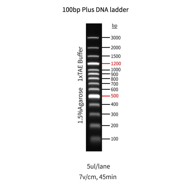
100bp plus DNA Ladder, 100-3000bp
Product description This product contains a DNA molecular weight marker consisting of 14 linear double-stranded DNA fragments at the following sizes: 100 bp; 200 bp; 300 bp; 400 bp; 500 bp; 600 bp; 700 bp; 800 bp; 900 bp; 1,000 bp; 1,200 bp; 1,500 bp; 2,000 bp; 3,000 bp. The reference bands are 500 bp and 1,200 bp, with a concentration of 120 ng/5 μL, while all other bands are at 50 ng/5 μL. The marker is supplied in 1×DNA Loading Buffer and is designed for agarose gel electrophoresis analysis of DNA bands. It is not recommended for polyacrylamide gel electrophoresis (PAGE). Specifications Product No. N132113S N132113M Size 100 T 10×100 T Components Component No. Component Name N132113S N132113M N132113-A 100 bp Plus DNA Ladder 500 μL 10×500 μL N132113-B 5×DNA Loading Buffer 1 mL 10×1 mL Shipping and Storage Store at room temperatures or at 2°C to 8°C, valid for half a year. Stor at -25℃ to -15℃, valid for one year. Avoid repeated freeze-thaw cycles. Notes 1. For optimal electrophoresis results: 1) Ensure thorough mixing of the product before use. 2) Replace the electrophoresis buffer promptly and use freshly prepared gels. 2. If smearing, blurred bands, or distortion occurs during electrophoresis: Dilute the sample with water before loading. For standard-width gel wells, dilute the sample 5-fold with water and load 8-10 μL. 3. When switching to a new stain or using agarose gels containing different stains: 1) Thoroughly clean the electrophoresis tank to avoid cross-contamination. 2) Replace with fresh electrophoresis buffer after cleaning. 4. For your safety and health, please wear a lab coat and disposable gloves. 5. For research use only! Instructions 1. Load 5 μL of the DNA ladder. For wide wells, increase the loading volume appropriately. 2. Use 1.5-2.0% agarose gels with a voltage of 4–10 V/cm in 0.5×TBE buffer or 1×TAE buffer. 3. Visualize DNA bands under UV light if stain the gel using solution-based staining methods with ethidium bromide (EB) or Arcegen Nucleic Acid Stain (Cat# N132109, non-toxic and UV-compatible).
$55.00 - $380.00

12000bp DNA Marker, 100-12000bp
Product description This product contains a DNA molecular weight marker consisting of 20 linear double-stranded DNA fragments at the following sizes: 12,000 bp; 8,000 bp; 6,000 bp; 5,000 bp; 4,000 bp; 3,000 bp; 2,500 bp; 2,000 bp; 1,500 bp; 1,200 bp; 1,000 bp; 900 bp; 800 bp; 700 bp; 600 bp; 500 bp; 400 bp; 300bp; 200 bp; 100 bp. The reference bands are 500 bp, 1,000 bp and 1,500 bp, with a concentration of 60 ng/5 μL, while all other bands are at 20 ng/5 μL. The marker is supplied in 1×DNA Loading Buffer and is designed for agarose gel electrophoresis analysis of DNA bands. It is not recommended for polyacrylamide gel electrophoresis (PAGE). Specifications Product No. N132120S N132120M Size 100 T 10×100 T Components Component No. Component Name N132120S N132120M N132120-A 12 kb DNA Marker 500 μL 10×500 μL N132120-B 5×DNA Loading Buffer 1 mL 10×1 mL Shipping and Storage Store at room temperatures or at 2°C to 8°C, valid for half a year. Stor at -25℃ to -15℃, valid for one year. Avoid repeated freeze-thaw cycles. Notes 1. For optimal electrophoresis results: 1) Ensure thorough mixing of the product before use. 2) Replace the electrophoresis buffer promptly and use freshly prepared gels. 2. If smearing, blurred bands, or distortion occurs during electrophoresis: Dilute the sample with water before loading. For standard-width gel wells, dilute the sample 5-fold with water and load 8-10 μL. 3. When switching to a new stain or using agarose gels containing different stains: 1) Thoroughly clean the electrophoresis tank to avoid cross-contamination. 2) Replace with fresh electrophoresis buffer after cleaning. 4. For your safety and health, please wear a lab coat and disposable gloves. 5. For research use only! Instructions 1. Load 5 μL of the DNA ladder. For wide wells, increase the loading volume appropriately. 2. Use 0.7-1.2% agarose gels with a voltage of 4–10 V/cm in 0.5×TBE buffer or 1×TAE buffer. 3. Visualize DNA bands under UV light if stain the gel using solution-based staining methods with ethidium bromide (EB) or Arcegen Nucleic Acid Stain (Cat# N132109, non-toxic and UV-compatible).
$55.00 - $3,000.00

15000bp DNA Marker, 250-15000bp
Product description This product contains a DNA molecular weight marker consisting of 7 linear double-stranded DNA fragments at the following sizes: 15,000 bp; 10,000 bp; 7,500 bp; 5,000 bp; 2,500 bp; 1,000 bp; 250 bp. The reference band is 2500 bp, with a concentration of 100 ng/5 μL. The marker is supplied in 1×DNA Loading Buffer and is designed for agarose gel electrophoresis analysis of DNA bands. It is not recommended for polyacrylamide gel electrophoresis (PAGE). Specifications Product No. N132121S N132121M Size 100 T 10×100 T Components Component No. Component Name N132121S N132121M N132121-A 15 kb DNA Marker 500 μL 10×500 μL N132121-B 5×DNA Loading Buffer 1 mL 10×1 mL Shipping and Storage Store at room temperatures or at 2°C to 8°C, valid for half a year. Stor at -25℃ to -15℃, valid for one year. Avoid repeated freeze-thaw cycles. Notes 1. For optimal electrophoresis results: 1) Ensure thorough mixing of the product before use. 2) Replace the electrophoresis buffer promptly and use freshly prepared gels. 2. If smearing, blurred bands, or distortion occurs during electrophoresis: Dilute the sample with water before loading. For standard-width gel wells, dilute the sample 5-fold with water and load 8-10 μL. 3. When switching to a new stain or using agarose gels containing different stains: 1) Thoroughly clean the electrophoresis tank to avoid cross-contamination. 2) Replace with fresh electrophoresis buffer after cleaning. 4. For your safety and health, please wear a lab coat and disposable gloves. 5. For research use only! Instructions 1. Load 5 μL of the DNA ladder. For wide wells, increase the loading volume appropriately. 2. Use 0.6% agarose gels with a voltage of 4–10 V/cm in 0.5×TBE buffer or 1×TAE buffer. 3. Visualize DNA bands under UV light if stain the gel using solution-based staining methods with ethidium bromide (EB) or Arcegen Nucleic Acid Stain (Cat# N132109, non-toxic and UV-compatible).
$55.00 - $380.00

1st Strand cDNA Synthesis SuperMix (gDNA digester plus, for qPCR)
Product description This product is a classic one-step cDNA synthesis reverse transcription ready-to-use premix from an RNA template. The core reverse transcriptase contained in the product has been effectively engineered to exhibit excellent thermal stability and template affinity, allowing it to tolerate reaction temperatures as high as 60°C. It is suitable for reverse transcription of RNA templates with complex secondary structures, low amounts of template, and low-copy genes. Additionally, this product contains a gDNA digestion component that can remove residual genomic DNA contamination from the RNA template, ensuring more reliable downstream results. The cDNA synthesized by this product is suitable for downstream qPCR applications. It is recommended to use it in combination with the Universal Multiplex qPCR Probe Premix (Cat#N132041), Universal qPCR Dye Premix (Cat#N132031), or the Traceable qPCR Dye Premix (Cat#N132034, Cat#N132035, Cat#N132036) for high-performance gene expression analysis. Specifications Catalog Number N132061E N132061S Specifications 10 T 100 T Components Catalog Number Component Name N132061E N132061S N132061-A RNase-free H2O 1 mL 2×1 mL N132061-B 5×gDNA Digester Mix 30 μL 320 μL N132061-C 4×cDNA Synthesis SuperMix 50 μL 500 μL 【Note】Component C of the 4×cDNA Synthesis SuperMix contains the gDNA Digester terminator. Storage Transport with ice packs. Store at -20℃. Valid for 18 months. Notes 1. Component B (5× gDNA Digester Mix) and Component C (4× cDNA Synthesis SuperMix) both contain high concentrations of glycerol. Before use, briefly centrifuge to collect the components at the bottom of the reaction tube. Then gently mix thoroughly by pipetting up and down, and accurately aspirate the required volume. 2. If the volume of the RNA template to be added is relatively large (e.g., more than 2 μL), ensure that the RNA is dissolved in water rather than TE buffer, as TE buffer may inhibit gDNA removal and reverse transcription. 3. The gDNA removal step can be omitted, and reverse transcription can be performed directly using Component C (4× cDNA Synthesis SuperMix). 4. The preparation of the reaction mixture should be carried out on ice to maintain stability. During the process, avoid contamination with RNase. 5. For your safety and health, please wear a lab coat and disposable gloves when operating. 6. For Research Use Only. Instructions 1. Removal of Residual Genomic DNA Prepare the following mixture in an RNase-free centrifuge tube and gently mix by pipetting. Incubate at 42°C for 2 minutes. Component Usage Amount RNase Free H2O To 15 μL 5×gDNA Digester Mix 3 μL Total RNA or mRNA RNA:10 pg-5 μg mRNA:10 pg-500 ng 【Note】For a 20 μL reverse transcription reaction system, it is recommended that the input amount of Total RNA does not exceed 1 μg. If the expression level of the target gene is low, up to 5 μg of Total RNA can be used. However, excessive RNA input may exceed the linear range of subsequent quantitative PCR. 2. Reverse Transcription Reaction Setup Directly add the 4× cDNA Synthesis SuperMix into the reaction tube from Step 1 and gently mix by pipetting. Component Usage Amount Step 1 Reaction Mixture 15 μL 4×cDNA Synthesis SuperMix 5 μL 3. Reverse Transcription Procedure Settings 1) Standard procedure Temperature Time 25℃ 5 min 55℃ 15 min 85℃ 5 min 【Note】The recommended reverse transcription temperature is 55℃. For templates with high GC content or complex structures, the reverse transcription temperature can be increased to 60℃. 2) Rapid Program [Suitable for templates with GC content ≤ 55% or simple templates] Temperature Time 55℃ 15 min 85℃ 5 sec 【Note】The reverse transcription product can be used immediately for qPCR reactions or stored short-term at -20°C. For long-term storage, it is recommended to aliquot and store at -80°C to avoid repeated freeze-thaw cycles.
$25.00 - $245.00

1× dsDNA HS Assay Kit (Ready to use)_N210302
The 1×dsDNA HS Assay Kit is a fast, sensitive, and accurate fluorescent quantification kit for double-stranded DNA (dsDNA). This kit offers high specificity for dsDNA and exhibits excellent linearity in the range of 0.2–100 ng. The assay is simple and convenient—just mix the sample with the working solution and read the fluorescence signal using a Qubit fluorometer or a fluorescence microplate reader. With its ease of use and reliable results, this kit is an ideal choice for high-throughput DNA quantification in next-generation sequencing (NGS) applications, such as input DNA quantification and DNA library quantification. The kit is also highly tolerant to common contaminants such as proteins and salts. Specifications Cat.No. N210302S / N210302M Size 100 T/500 T Components Components No. Name Concentration N210302S N210302M N210302-A 1×dsDNA HS Working Solution 1×concentrate in DMSO 50 mL 250 mL N210302-B dsDNA Standard 1 0 ng/μL in TE buffer 1 mL 5×1 mL N210302-C dsDNA Standard 2 10 ng/μL in TE buffer 1 mL 5×1 mL Storage Shipped on ice packs. Store at 2–8 °C in the dark, valid for 1 year. Documents: Manual
$85.00 - $265.00

2000bp DNA Marker, 100-2000bp
Product description This product contains a DNA molecular weight marker consisting of 6 linear double-stranded DNA fragments at the following sizes: 2,000 bp; 1,000 bp; 750 bp; 500 bp; 250 bp; 100 bp. The reference band is 750 bp, with a concentration of 125 ng/5 μL, while all other bands are at 50 ng/5 μL. The marker is supplied in 1×DNA Loading Buffer and is designed for agarose gel electrophoresis analysis of DNA bands. It is not recommended for polyacrylamide gel electrophoresis (PAGE). Specifications Product No. N132117S N132117M Size 100 T 10×100 T Components Component No. Component Name N132117S N132117M N132117-A 2 kb DNA Marker 500 μL 10×500 μL N132117-B 5×DNA Loading Buffer 1 mL 10×1 mL Shipping and Storage Store at room temperatures or at 2°C to 8°C, valid for half a year. Stor at -25℃ to -15℃, valid for one year. Avoid repeated freeze-thaw cycles. Notes 1. For optimal electrophoresis results: 1) Ensure thorough mixing of the product before use. 2) Replace the electrophoresis buffer promptly and use freshly prepared gels. 2. If smearing, blurred bands, or distortion occurs during electrophoresis: Dilute the sample with water before loading. For standard-width gel wells, dilute the sample 5-fold with water and load 8-10 μL. 3. When switching to a new stain or using agarose gels containing different stains: 1) Thoroughly clean the electrophoresis tank to avoid cross-contamination. 2) Replace with fresh electrophoresis buffer after cleaning. 4. For your safety and health, please wear a lab coat and disposable gloves. 5. For research use only! Instructions 1. Load 5 μL of the DNA ladder. For wide wells, increase the loading volume appropriately. 2. Use 1.0-2.0% agarose gels with a voltage of 4–10 V/cm in 0.5×TBE buffer or 1×TAE buffer. 3. Visualize DNA bands under UV light if stain the gel using solution-based staining methods with ethidium bromide (EB) or Arcegen Nucleic Acid Stain (Cat# N132109, non-toxic and UV-compatible).
$55.00 - $385.00

200bp DNA Ladder, 200-5000bp
Product description This product contains a DNA molecular weight marker consisting of 12 linear double-stranded DNA fragments at the following sizes: 200 bp; 400 bp; 600 bp; 800 bp; 1,000 bp; 1,200 bp; 1,400 bp; 1,600 bp; 1,800 bp; 2,000 bp; 3,000 bp; 5,000 bp. The reference band is 1,200 bp, with a concentration of 100 ng/5 μL, while all other bands are at 40 ng/5 μL. The marker is supplied in 1×DNA Loading Buffer and is designed for agarose gel electrophoresis analysis of DNA bands. It is not recommended for polyacrylamide gel electrophoresis (PAGE). Specifications Product No. N132114S N132114M Size 100 T 10×100 T Components Component No. Component Name N132114S N132114M N132114-A 200 bp DNA Ladder 500 μL 10×500 μL N132114-B 5×DNA Loading Buffer 1 mL 10×1 mL Shipping and Storage Store at room temperatures or at 2°C to 8°C, valid for half a year. Stor at -25℃ to -15℃, valid for one year. Avoid repeated freeze-thaw cycles. Notes 1. For optimal electrophoresis results: 1) Ensure thorough mixing of the product before use. 2) Replace the electrophoresis buffer promptly and use freshly prepared gels. 2. If smearing, blurred bands, or distortion occurs during electrophoresis: Dilute the sample with water before loading. For standard-width gel wells, dilute the sample 5-fold with water and load 8-10 μL. 3. When switching to a new stain or using agarose gels containing different stains: 1) Thoroughly clean the electrophoresis tank to avoid cross-contamination. 2) Replace with fresh electrophoresis buffer after cleaning. 4. For your safety and health, please wear a lab coat and disposable gloves. 5. For research use only! Instructions 1. Load 5 μL of the DNA ladder. For wide wells, increase the loading volume appropriately. 2. Use 1.5-2.0% agarose gels with a voltage of 4–10 V/cm in 0.5×TBE buffer or 1×TAE buffer. 3. Visualize DNA bands under UV light if stain the gel using solution-based staining methods with ethidium bromide (EB) or Arcegen Nucleic Acid Stain (Cat# N132109, non-toxic and UV-compatible).
$55.00 - $385.00

2× Color qPCR Fluore Green Master Mix (High Rox)
Product description The 2×Color qPCR Fluore Green Master Mix (High Rox) is a 2× real-time quantitative PCR amplification premix, characterized by high fluorescence intensity, high sensitivity, strong specificity, and high amplification yield. The core component, Taq DNA polymerase, utilizes an antibody-mediated hot-start mechanism, which effectively inhibits non-specific amplification caused by primer annealing during sample preparation. Additionally, the mix contains factors that enhance PCR amplification efficiency and promote balanced amplification across a wide range of GC content (30-70%), ensuring good linearity in quantitative PCR over a broad quantification range. This product employs a color-changing reaction based on the mixture of different dyes to monitor pipetting operations, effectively reducing the occurrence of pipetting errors. Specifications Catalog Number N132036E N132036S Specifications 50 T(20 μL/rxn) 250 T(20 μL/rxn) Components Component Identification Component Name N132036E N132036S N132036-A 2×Color qPCR Master Mix (High Rox) 1 mL 5×1 mL N132036-B 10×Dilution Buffer 1 mL 1 mL Storage Store at -25 to -15℃ in the dark. Valid for 1 year. Notes 1. For your safety and health, please wear a lab coat and disposable gloves when operating. 2. After thawing, the Master Mix may appear with flocculent or white precipitates. Hold it in hand and gently dissolve by slowly inverting it up and down until the solution is clear. This does not affect the performance of the reagent. 3. Before use, invert the Master Mix gently to mix. Do not vortex to avoid generating bubbles, which may affect the quantification results. The Master Mix can be used after mixing and brief centrifugation. During pipetting, gently aspirate and dispense. If bubbles form in the Master Mix due to improper operation, centrifuge it again before use. 4. Due to the extremely high sensitivity of this product, it is susceptible to aerosol contamination in the air. Therefore, when preparing the reaction mixture, please do it in a laminar flow cabinet. Use sterile pipette tips and reaction tubes during preparation. Laboratories with the conditions are recommended to use dedicated pipettes and filter-tipped pipette tips. 5. It is recommended to use our company's cDNA synthesis kit (Cat#N132062) to effectively remove residual genomic material from RNA samples. 6. For Research Use Only. Instructions 1. Template Processing The 2×Color qPCR Fluore Green Master Mix (No Rox) contains a blue dye, and the 10×Dilution Buffer is a dedicated yellow concentrated template dilution solution. During use, if template tracing is required, dilute it to 1X as the template dilution solution and then proceed with qPCR detection. If template tracing is not required, simply do not use the Dilution Buffer. Template Types Usage of 10× Dilution Buffer Final Concentration cDNA solution, dissolved plasmid, genome If you need to dilute the template, first dilute the template to the desired concentration with sterile ultrapure water. Then, add 1 μL of 10×Dilution Buffer to every 9 μL of the diluted template. 1× DNA Powder Dilute the 10×Dilution Buffer to 1× using sterile ultrapure water. Dissolve the corresponding DNA powder using an appropriate volume of 1× Dilution Buffer as the solvent. 1× 【Note】In actual use, other methods can also be employed as needed, as long as the final concentration of Dilution Buffer in the template is 1×. 2. Recommended qPCR Reaction System Component Volume(μL)*** Volume(μL)*** Final concentration 2×miRNA qPCR Master Mix 25 10 1× Forward Primer(self-provided)* 1 0.4 0.2 μM Reverse Primer(10 μM)* 1 0.4 0.2 μM cDNA ** x x - RNase-free H2O Up to 50 Up to 20 - 【Note】*The typical final concentration of primers is 0.2 μM, but it can also be adjusted between 0.1-1.0 μM depending on the situation. **If the template is undiluted cDNA stock solution, the volume used should not exceed 1/10 of the total qPCR reaction volume. The optimal amount of template added should result in Ct values between 20-30 cycles for amplification. ***A reaction volume of 20 μL or 50 μL is recommended to ensure the effectiveness and reproducibility of target gene amplification. Mix thoroughly before running the reaction to avoid excessive bubbles from vigorous shaking. 3. Reaction Program Recycling procedure Temperature Time Cycle Number Recycling procedure 95℃ 2 min 1 Pre-denaturation 95℃ 10 sec 40 Denaturation 60℃ 30 sec** Melting Curve Default Settings 1 【Note】*Annealing Temperature and Time: Please adjust according to the primer Tm value and the length of the target gene. **Fluorescence Signal Acquisition: Do not forget to enable fluorescence signal acquisition. Set up the experimental procedure according to the user manual of the instrument. ***Melting Curve: The default program of the instrument can usually be used. 4. Primer Design Guidelines 1) The recommended primer length is around 25 bp. The optimal amplicon size is 150 bp, but it can be chosen within the range of 100 bp to 300 bp. 2) The Tm values of the forward and reverse primers should not differ by more than 2℃. A Tm value between 60℃ and 65℃ is preferred. 3) The base distribution in the primer should be uniform, avoiding four consecutive identical bases. The GC content should be controlled around 50%. The last base at the 3’ end should preferably be G or C. 4) Avoid complementary sequences of more than three bases within a primer or between the forward and reverse primers. 5) The specificity of the primers should be verified using the NCBI BLAST program. Avoid having more than two non-specific complementary bases at the 3’ end of the primers. 6) The designed primers need to be tested for amplification efficiency. Only primers with the same amplification efficiency should be used for quantitative comparative analysis 5. Compatible Instruments This product contains a pre-mixed ROX Reference Dye 1 (high concentration) for correcting well-to-well fluorescence signal errors, and is suitable for the following real-time fluorescent quantitative PCR instruments: Applied Biosystems 5700, 7000, 7300, 7700, 7900, 7900HT, 7900HT Fast, StepOne, StepOnePlus; And other real-time fluorescent quantitative PCR instruments that require the addition of high-concentration ROX Reference Dye.
$60.00 - $240.00

2× Color qPCR Fluore Green Master Mix (Low Rox)
Product description The 2×Color qPCR Fluore Green Master Mix (Low Rox) is a 2× real-time quantitative PCR amplification premix, characterized by high fluorescence intensity, high sensitivity, strong specificity, and high amplification yield. The core component, Taq DNA polymerase, utilizes an antibody-mediated hot-start mechanism, which effectively inhibits non-specific amplification caused by primer annealing during sample preparation. Additionally, the mix contains factors that enhance PCR amplification efficiency and promote balanced amplification across a wide range of GC content (30-70%), ensuring good linearity in quantitative PCR over a broad quantification range. This product employs a color-changing reaction based on the mixture of different dyes to monitor pipetting operations, effectively reducing the occurrence of pipetting errors. Specifications Catalog Number N132035E N132035S Specifications 50 T(20 μL/rxn) 250 T(20 μL/rxn) Components Component Identification Component Name N132034E N132034S N132035-A 2×Color qPCR Master Mix (Low Rox) 1 mL 5×1 mL N132035-B 10×Dilution Buffer 1 mL 1 mL Storage Store at -25 to -15℃ in the dark. Valid for 1 year. Notes 1. For your safety and health, please wear a lab coat and disposable gloves when operating. 2. After thawing, the Master Mix may appear with flocculent or white precipitates. Hold it in hand and gently dissolve by slowly inverting it up and down until the solution is clear. This does not affect the performance of the reagent. 3. Before use, invert the Master Mix gently to mix. Do not vortex to avoid generating bubbles, which may affect the quantification results. The Master Mix can be used after mixing and brief centrifugation. During pipetting, gently aspirate and dispense. If bubbles form in the Master Mix due to improper operation, centrifuge it again before use. 4. Due to the extremely high sensitivity of this product, it is susceptible to aerosol contamination in the air. Therefore, when preparing the reaction mixture, please do it in a laminar flow cabinet. Use sterile pipette tips and reaction tubes during preparation. Laboratories with the conditions are recommended to use dedicated pipettes and filter-tipped pipette tips. 5. It is recommended to use our company's cDNA synthesis kit (Cat#N132062) to effectively remove residual genomic material from RNA samples. 6. For Research Use Only. Instructions 1. Template Processing The 2×Color qPCR Fluore Green Master Mix (No Rox) contains a blue dye, and the 10×Dilution Buffer is a dedicated yellow concentrated template dilution solution. During use, if template tracing is required, dilute it to 1X as the template dilution solution and then proceed with qPCR detection. If template tracing is not required, simply do not use the Dilution Buffer. Template Types Usage of 10× Dilution Buffer Final Concentration cDNA solution, dissolved plasmid, genome If you need to dilute the template, first dilute the template to the desired concentration with sterile ultrapure water. Then, add 1 μL of 10×Dilution Buffer to every 9 μL of the diluted template. 1× DNA Powder Dilute the 10×Dilution Buffer to 1× using sterile ultrapure water. Dissolve the corresponding DNA powder using an appropriate volume of 1× Dilution Buffer as the solvent. 1× 【Note】In actual use, other methods can also be employed as needed, as long as the final concentration of Dilution Buffer in the template is 1×. 2. Recommended qPCR Reaction System Component Volume(μL)*** Volume(μL)*** Final concentration 2×miRNA qPCR Master Mix 25 10 1× Forward Primer(self-provided)* 1 0.4 0.2 μM Reverse Primer(10 μM)* 1 0.4 0.2 μM cDNA ** x x - RNase-free H2O Up to 50 Up to 20 - 【Note】*The typical final concentration of primers is 0.2 μM, but it can also be adjusted between 0.1-1.0 μM depending on the situation. **If the template is undiluted cDNA stock solution, the volume used should not exceed 1/10 of the total qPCR reaction volume. The optimal amount of template added should result in Ct values between 20-30 cycles for amplification. ***A reaction volume of 20 μL or 50 μL is recommended to ensure the effectiveness and reproducibility of target gene amplification. Mix thoroughly before running the reaction to avoid excessive bubbles from vigorous shaking. 3. Reaction Program Recycling procedure Temperature Time Cycle Number Recycling procedure 95℃ 2 min 1 Pre-denaturation 95℃ 10 sec 40 Denaturation 60℃ 30 sec** Melting Curve Default Settings 1 【Note】*Annealing Temperature and Time: Please adjust according to the primer Tm value and the length of the target gene. **Fluorescence Signal Acquisition: Do not forget to enable fluorescence signal acquisition. Set up the experimental procedure according to the user manual of the instrument. ***Melting Curve: The default program of the instrument can usually be used. 4. Primer Design Guidelines 1) The recommended primer length is around 25 bp. The optimal amplicon size is 150 bp, but it can be chosen within the range of 100 bp to 300 bp. 2) The Tm values of the forward and reverse primers should not differ by more than 2℃. A Tm value between 60℃ and 65℃ is preferred. 3) The base distribution in the primer should be uniform, avoiding four consecutive identical bases. The GC content should be controlled around 50%. The last base at the 3’ end should preferably be G or C. 4) Avoid complementary sequences of more than three bases within a primer or between the forward and reverse primers. 5) The specificity of the primers should be verified using the NCBI BLAST program. Avoid having more than two non-specific complementary bases at the 3’ end of the primers. 6) The designed primers need to be tested for amplification efficiency. Only primers with the same amplification efficiency should be used for quantitative comparative analysis 5. Compatible Instruments This product contains a pre-mixed ROX Reference Dye 2 (low concentration) for correcting well-to-well fluorescence signal errors, and is suitable for the following real-time fluorescent quantitative PCR instruments: Applied Biosystems 7500, 7500 Fast, ViiA7; Stratagene MX4000, MX3005P, MX3000P; And other real-time fluorescent quantitative PCR instruments that require the addition of low-concentration ROX Reference Dye.
$60.00 - $240.00
-
2× Color qPCR Fluore Green Master Mix (No Rox)
Product description The 2×Color qPCR Fluore Green Master Mix (No Rox) is a 2× real-time quantitative PCR amplification premix, characterized by high amplification yield, high fluorescence intensity, high sensitivity, and strong specificity. The core component, Taq DNA polymerase, utilizes an antibody-mediated hot-start mechanism, which effectively inhibits non-specific amplification caused by primer annealing during sample preparation. Additionally, the mix contains factors that enhance PCR amplification efficiency and promote balanced amplification across a wide range of GC content (30-70%), ensuring good linearity in quantitative PCR over a broad quantification range. This product employs a color-changing reaction based on the mixture of different dyes to monitor pipetting operations, effectively reducing the occurrence of pipetting errors. Specifications Catalog Number N132034E N132034S Specifications 50 T(20 μL/rxn) 250 T(20 μL/rxn) Components Component Identification Component Name N132034E N132034S N132034-A 2×Color qPCR Master Mix (No Rox) 1 mL 5×1 mL N132034-B 10×Dilution Buffer 1 mL 1 mL Storage Store at -25 to -15℃ in the dark. Valid for 1 year. Notes 1. For your safety and health, please wear a lab coat and disposable gloves when operating. 2. After thawing, the Master Mix may appear with flocculent or white precipitates. Hold it in hand and gently dissolve by slowly inverting it up and down until the solution is clear. This does not affect the performance of the reagent. 3. Before use, invert the Master Mix gently to mix. Do not vortex to avoid generating bubbles, which may affect the quantification results. The Master Mix can be used after mixing and brief centrifugation. During pipetting, gently aspirate and dispense. If bubbles form in the Master Mix due to improper operation, centrifuge it again before use. 4. Due to the extremely high sensitivity of this product, it is susceptible to aerosol contamination in the air. Therefore, when preparing the reaction mixture, please do it in a laminar flow cabinet. Use sterile pipette tips and reaction tubes during preparation. Laboratories with the conditions are recommended to use dedicated pipettes and filter-tipped pipette tips. 5. It is recommended to use our company's cDNA synthesis kit (Cat#N132062) to effectively remove residual genomic material from RNA samples. 6. For Research Use Only. Instructions 1. Template Processing The 2×Color qPCR Fluore Green Master Mix (No Rox) contains a blue dye, and the 10×Dilution Buffer is a dedicated yellow concentrated template dilution solution. During use, if template tracing is required, dilute it to 1X as the template dilution solution and then proceed with qPCR detection. If template tracing is not required, simply do not use the Dilution Buffer. Template Types Usage of 10× Dilution Buffer Final Concentration cDNA solution, dissolved plasmid, genome If you need to dilute the template, first dilute the template to the desired concentration with sterile ultrapure water. Then, add 1 μL of 10×Dilution Buffer to every 9 μL of the diluted template. 1× DNA Powder Dilute the 10×Dilution Buffer to 1× using sterile ultrapure water. Dissolve the corresponding DNA powder using an appropriate volume of 1× Dilution Buffer as the solvent. 1× 【Note】In actual use, other methods can also be employed as needed, as long as the final concentration of Dilution Buffer in the template is 1×. 2. Recommended qPCR Reaction System Component Volume(μL)*** Volume(μL)*** Final concentration 2×miRNA qPCR Master Mix 25 10 1× Forward Primer(self-provided)* 1 0.4 0.2 μM Reverse Primer(10 μM)* 1 0.4 0.2 μM cDNA ** x x - RNase-free H2O Up to 50 Up to 20 - 【Note】*The typical final concentration of primers is 0.2 μM, but it can also be adjusted between 0.1-1.0 μM depending on the situation. **If the template is undiluted cDNA stock solution, the volume used should not exceed 1/10 of the total qPCR reaction volume. The optimal amount of template added should result in Ct values between 20-30 cycles for amplification. ***A reaction volume of 20 μL or 50 μL is recommended to ensure the effectiveness and reproducibility of target gene amplification. Mix thoroughly before running the reaction to avoid excessive bubbles from vigorous shaking. 3. Reaction Program Recycling procedure Temperature Time Cycle Number Recycling procedure 95℃ 2 min 1 Pre-denaturation 95℃ 10 sec 40 Denaturation 60℃ 30 sec** Melting Curve Default Settings 1 【Note】*Annealing Temperature and Time: Please adjust according to the primer Tm value and the length of the target gene. **Fluorescence Signal Acquisition: Do not forget to enable fluorescence signal acquisition. Set up the experimental procedure according to the user manual of the instrument. ***Melting Curve: The default program of the instrument can usually be used. 4. Primer Design Guidelines 1) The recommended primer length is around 25 bp. The optimal amplicon size is 150 bp, but it can be chosen within the range of 100 bp to 300 bp. 2) The Tm values of the forward and reverse primers should not differ by more than 2℃. A Tm value between 60℃ and 65℃ is preferred. 3) The base distribution in the primer should be uniform, avoiding four consecutive identical bases. The GC content should be controlled around 50%. The last base at the 3’ end should preferably be G or C. 4) Avoid complementary sequences of more than three bases within a primer or between the forward and reverse primers. 5) The specificity of the primers should be verified using the NCBI BLAST program. Avoid having more than two non-specific complementary bases at the 3’ end of the primers. 6) The designed primers need to be tested for amplification efficiency. Only primers with the same amplification efficiency should be used for quantitative comparative analysis 5. Compatible Instruments This product does not include ROX Reference Dye for correcting well-to-well fluorescence signal errors. It is compatible with the following real-time fluorescent quantitative PCR instruments: Bio-Rad CFX96, CFX384, iCycler iQ, iQ5, MyiQ; MiniOpticon, Opticon, Opticon 2, Chromo4; Cepheid SmartCycler; Eppendorf Mastercycler ep realplex, realplex 2 s; Illumina Eco qPCR; Qiagen/Corbett Rotor-Gene Q, Rotor-Gene 3000, Rotor-Gene 6000; Roche Applied Science LightCycler 480; Thermo Scientific PikoReal Cycler; and other real-time fluorescent quantitative PCR instruments that do not require the addition of ROX Reference Dye.Dye-based qPCRdye-based
$60.00 - $240.00

2× High-Fidelity Fast PCR Master Mix (With Dye)
Product description This kit is a ready-to-use 2×pre-mixed solution containing High-Fidelity DNA Polymerase, dNTPs, and an optimized buffer system, which contains pre-added electrophoresis indicators. The pre-mix contains pre-added electrophoresis indicator, PCR products can be directly electrophoresed, the amplification products are flat ends. 2×High-Fidelity Fast PCR Master Mix (With Dye) has the advantages of quick and easy, high sensitivity, high specificity, good stability, etc., the reaction system can be added with only the primers and templates. In addition, the product also contains a specific protective agent, so that the premix can still maintain stable activity after repeated freezing and thawing. Components Components No. N132016S N132016M Size 1 mL 5×1 mL Shipping and Storage Dry ice shipping. -15℃ ~ -25℃ storage, valid for one year. Notes 1. This product is for research use only. 2. Please operate with lab coats and disposable gloves, for your safety. Instructions 1. Recommended PCR reaction systems. Table 1 PCR reaction system Components Volume(μL) Final concentration 2×High-Fidelity Fast PCR Master Mix (With Dye) 25 1× Template** x - Forward Primer(10 μmol/L)*** 2 0.4 μmol/L Reverse Primer(10 μmol/L) 2 0.4 μmol/L ddH2O Up to 50 - 【Note】: a. *In 1× premixes containing 2 mM Mg2+ and 200 μM dNTPs. b. **Recommended range 10-200 ng, cDNA sample upload volume range not more than 1/10 of the reaction system, recommended 1-2.5 μL. c. ***The final primer concentration in the PCR reaction system ranges from 0.2-1 μM, and 0.4 μM is recommended. 2. Reaction program Table 2 PCR reaction program Cycle step Temp. Time Cycles Initial denaturation 98℃ 30 sec 1 Denaturation 98℃ 10 sec 30-35 Annealing* 60℃ 5 sec Extension** 72℃ 5-10 sec/kb Final extension 72℃ 2 min 1 【Note】: 1. *Recommended temperature: 60°C, a temperature gradient can be set up to find the optimal temperature for primer annealing. The recommended annealing time is set to 5 sec and can be adjusted from 5-30 sec. Too long annealing time may result in diffuse amplification products on the gel. 2. **Extension time: Recommended 5 sec/kb, can also be extended to 10 sec/kb as needed.
$30.00 - $385.00
-
2× High-Fidelity PCR Master Mix (With Dye)
Product description This kit is a ready-to-use 2×pre-mixed solution containing High-Fidelity DNA Polymerase, dNTPs, and an optimized buffer system. The mix includes two monoclonal antibodies that inhibit polymerase activity and 3’→5’exonuclease activity at room temperature, enabling highly specific Hot Start PCR. The addition of elongation factors allows for long-fragment amplification, with target fragments up to 13 kb. This enzyme exhibits 5’→3’ DNA polymerase activity and 3’→5’ exonuclease activity, with a fidelity 83 times that of Taq DNA polymerase and 9 times that of ordinary Pfu DNA polymerase. It is suitable for amplifying complex templates, producing blunt-end PCR products. The kit offers advantages such as simplicity, high sensitivity, strong specificity, and excellent stability. The reaction system only requires the addition of primers and template, and a two-step PCR protocol can be used to simplify the procedure and save time. The product includes electrophoresis tracking dye, allowing direct loading of PCR products for gel analysis. Additionally, it contains a special protective agent that ensures stable activity even after repeated freeze-thaw cycles. Components Components N132014S N132014M N132014L 2× High-Fidelity PCR Master Mix (With Dye) 1 mL 5×1 mL 100×1 mL Shipping and Storage Dry ice shipping. -15℃ ~ -25℃ storage, valid for one year. Notes 1. This product is for research use only. 2. Please operate with lab coats and disposable gloves, for your safety. Instructions Recommended PCR reaction systems. Table 1 PCR reaction system Components Volume(μL) Final concentration 2× High-Fidelity PCR Master Mix (With Dye)* 25 1× Template*** x - Forward Primer(10 μmol/L)** 2 0.4 μmol/L Reverse Primer(10 μmol/L)** 2 0.4 μmol/L ddH2O Up to 50 - 【Note】: 1) *The 1× final concentration of the mix contains 1 U/50 μL of polymerase, 2 mmol/L Mg2+, and 200 μmol/L dNTPs. 2) **The recommended final concentration of primers in the PCR reaction is 0.2-1 μmol/L, with 0.4 μmol/L preferred. 3) ***Recommended template amounts for a 25 μL reaction system: Table 2 Recommended template amounts Template Type Fragment Size (1 kb-10 kb) Genomic DNA 50 ng-200 ng Plasmid or Viral DNA 10 pg-20 ng cDNA 1-2.5 μL (not exceeding 1/10 of the total PCR volume) Reaction program. 1)Two-Step Protocol (Preferred): Cycle step Temp. Time Cycles Initial denaturation 98℃ 3 min 1 Denaturation 98℃ 10 sec 30-35 Extension 68℃ 30 sec/kb Final extension 72℃ 5 min 1 2)Three-Step Protocol (Conventional): Cycle step Temp. Time Cycles Initial denaturation* 98℃ 3 min 1 Denaturation 98℃ 10 sec 30-35 Annealing** 60℃ 20 sec Extension*** 72℃ 30 sec/kb Final extension 72℃ 5 min 1 【Note】: a. * Initial Denaturation: Recommended at 98°C for 3 min (5-10 min for high-GC templates). b. **Annealing: Recommended at 60°C for 20 sec (adjustable between 10-30 sec). Prolonged annealing may cause smearing on gels. c. ***Extension: Recommended at 72°C for 30 sec/kb (up to 60 sec/kb for complex templates). 3)Gradient Annealing Protocol (Recommended for Difficult Targets): Cycle step Temp. Time Cycles Initial denaturation 98℃ 3 min 1 Denaturation 98℃ 10 sec 15 (decrease by 1° C per cycle) Gradient Annealing 70-55°C 20 sec Extension 72℃ 30 sec/kb Denaturation 98°C 10 sec 20 Annealing 55°C 20 sec Extension 72°C 30 sec/kb Final extension 72℃ 5 min 1 4)Characteristics of Different PCR Protocols: Protocol Type Speed Specificity PCR Yield Detection Rate Two-Step Fastest High Medium High Three-Step Medium Medium Highest Medium Gradient Annealing Slow High Medium High
$15.00 - $3,665.00

2× HotStart Fast PCR Master Mix (With Dye)
Product description 2× HotStart PCR Master Mix contains heat-stabilized Taq DNA Polymerase modified with antibodies, adds strong elongation factor and optimized buffer system, and has super high amplification efficiency. It is very suitable for PCR amplification of most colonies such as E. coli, Agrobacterium, and yeast. The extension speed of amplification of complex templates within 3 kb can reach 1 sec/kb, 3-6 kb to 3 sec/kb, 6-10 kb to 5 sec/kb and 10 kb to 10 sec/kb; the amplification speed of simple templates such as plasmid within 6 kb can reach 1 sec/kb, which can greatly save PCR reaction time. Meanwhile, Mix contains dNTPs, Mg2+, and it can be amplified only with adding primers and template. In addition, Mix contains red tracer dye, which can be used by electrophoresis directly after the end of the reaction. The protective agent added to the system enables the product to maintain the stable activity after repeated freezing and thawing. The 3′ end of the PCR product bands A and can be easily cloned into the T vector. Components Components No. N132001E N132001S N132001M N132001L Size 1 mL 5×1 mL 50×1 mL 100×1 mL Specifications Product specification Master Mix Concentration 2× Hot Start Built-in Hot Start Overhang 3 '-A Reaction speed Rapid Size (Final Product) Up to 15 kb Conditions for transportation Dry ice Shipping and Storage Dry ice shipping. -15℃ ~ -25℃ storage, valid for two years. Notes 1. For your safety and health, please wear lab coats and disposable gloves for operation. 2. This product is for research use ONLY! Instructions 1. Reaction System Table 1 Reaction system Components Size (μL) Size (μL) Final concentration 2×HotStart Fast PCR Master Mix (With Dye)* 25 12.5 1× Template DNA** suitable suitable - Forward primer (10 μmol/L) 2 1 0.4-0.5 μM Reverse primer (10 μmol/L) 2 1 0.4-0.5 μM ddH2O to 50 to 25 - 【Note】: 1) *The 2×Mix contains 2 mM Mg2+ and 200 μM dNTPs, thawed thoroughly before use. 2) **E. coli and Agrobacterium can directly absorb bacterial liquid or pick bacteria samples; It is recommended that the yeast liquid and 3) **Strain be boiled for 5 min, then put into the -80℃ refrigerator for 3 min, and then serve the sample after thawing. Note that the bacterial liquid sample should be shaken and mixed before sampling, in which the recommended amount of bacterial liquid sample is 2-4 μL (0.5-0.8 OD600). Recommended usage of the different templates: Table 2 Recommended usage of different templates Type of template Segment usage range (25 μL reaction system) Genomic DNA 10–1,000 ng Plasmid or λDNA 0.5-50 ng E. coli bacteria solution 0.5-0.8 OD600 【Note】: ***The range of final primer concentration in PCR reaction system is 0.2-1 μM, and 0.4 μM is recommended. 2. Amplification Protocol Table 3 Amplification protocol Cycle steps Temperature (°C) Time Cycles Predenaturation 95 3 min 1 Denaturation 95 15 sec 30-35 Annealing* 60 20 sec Extension** 72 1-10 sec/kb Final extension 72 5 min 1 【Note】: a. *Recommended annealing temperature: 60°C, you can also set up a temperature gradient according to your own needs to explore the optimal temperature for primer annealing. The recommended annealing time is set to 20 sec, which can be adjusted within 10-30 sec. Too long annealing time may lead to dispersion of the amplified product on the glue. b. **Extension speed: 1sec/kbfor complex templates such as genomes and E. coli within 3 kb, 3 sec/kb for complex templates within 6 kb, 5 sec/kb for most complex templates within 10 kb, and 10 sec/kb for complex template fragments over 10 kb. For simple templates, such as plasmids less than 6 kb, set 1 sec/kb; for simple templates, such as 6-10 kb plasmids, set 3 sec/kb; and for simple templates, such as plasmids larger than 10 kb, set 5-10 sec/kb. If it is necessary to increase production, the extension time can be extended appropriately, and should not exceed 30 sec/kb.
$45.00 - $2,970.00

2× HotStart Genotyping PCR Master Mix (With Dye)
Product description The kit is a ready-to-use PCR premixed solution containing HotStart Taq DNA Polymerase, dNTPs, and an optimized buffering system. Simply add primers and template for amplification, which greatly simplifies the experimental procedure and allows for high-throughput operations, enhancing the reproducibility of results. The hotStart Taq DNA Polymerase is a ligand-modified thermostable Taq DNA Polymerase, with a ligand that modulates the activity of the DNA polymerase in response to temperature changes. The enzyme activity is completely sealed at room temperature and is only released after heating at 95℃. The activation time for HotStart DNA Polymerase requires only 2-3 min and is compatible with existing PCR protocols. This product prevents non-specific amplification during sample preparation and reaction heating stages, effectively enabling genotyping experiments. Components Name N132004E N132004S N132004M N132004L 2×HotStart Genotyping PCR Master Mix (With Dye) 1 mL 5×1 mL 50×1 mL 100×1 mL Specifications Concentration 2× Hot Start Built-in Hot Start Overhang 3 '-A Polymerase Taq DNA Polymerase Reaction speed Standard Product Type PCR Master Mix (2x) Shipping and Storage Dry ice shipping. -15℃ ~ -25℃ storage, valid for two years. Applications This product is primarily applied to the genotyping of mice. Notes 1. For your safety and health, please wear lab coats and disposable gloves for operation. 2. This product is for research use ONLY!This product is for research use ONLY! Instructions 1. Reaction System Table 1 Reaction system Components Size (μL) Final concentration 2×HotStart Genotyping PCR Master Mix (With Dye) 25 1× Template DNA suitable - Forward primer (10 μmol/L) 2 0.4 μM Reverse primer (10 μmol/L) 2 0.4 μM ddH2O to 50 - Recommended usage of the different templates: Table 2 Recommended usage of different templates Type of template Segment usage range (50 μL reaction system) Genomic DNA 50–100 ng Plasmid DNA 0.1-20 ng cDNA 1-5 μL (Do not exceed 1/10 of the reaction system) 2. Amplification Protocol Table 3 Amplification protocol Cycle steps Temperature (°C) Time Cycles Predenaturation 95 5 min 1 Denaturation 95 30 sec 35 Annealing 50-60 30 sec Extension 72 30-60 sec/kb Final extension 72 10 min 1 【Note】: a. *Recommended Annealing Temperature and Time: The annealing temperature is recommended to be set at 50-60℃. The annealing time is recommended to be set at 30 sec, with adjustments possible within the range of 20-30 sec. Depending on the need, a temperature gradient can be established to explore the optimal annealing temperature and time for primer annealing. b. **Extension Temperature and Time: The temperature is recommended to be 72℃. The time is recommended to be 30-60 sec/kb. c. **PCR Amplification Products: Please store the PCR amplification products at -20℃ to prevent DNA degradation.
$35.00 - $1,550.00
-
2× miRNA qPCR Fluore Green Master Mix
Product description The 2×miRNA qPCR Fluore Green Master Mix is a new-generation premixed fluorescent quantitative PCR detection reagent specifically developed for miRNA quantification. It contains a special ROX Passive Reference Dye, which is compatible with all qPCR instruments without the need to adjust the ROX concentration for different instruments. The DNA Polymerase used is a chemically modified hot-start polymerase, combined with a special buffer system, which enhances the specificity of the reaction, improves sensitivity, and allows for accurate quantification over a broader range. This product must be used in conjunction with our miRNA First-Strand cDNA Synthesis Reverse Transcription Kit (Poly(A) tailing method, for qPCR) (Cat#N132068) to obtain the final experimental results. Specifications Catalog Number N132039E N132039S Specifications 100 T(20 μL/rxn) 500T(20 μL/rxn) Components Component Identification Component Name N132036E N132036S N132039-A 2×miRNA qPCR Master Mix 1 mL 5×1 mL N132039-B RNase-free H2O 2×1 mL 5×1 mL Storage Ice bag transportation. Store at -20℃ in the dark. Valid for 12 months. Notes 1. Thaw the components completely and gently mix them before use. 2. During the experiment, use contamination-free consumables whenever possible to avoid contamination. 3. Avoid repeated freeze-thaw cycles for this product. Protect from strong light during preparation. 4. For your safety and health, please wear a lab coat and disposable gloves when operating. 5. This product is for research use only. Instructions 1. Reaction Preparation 1) Place the reagents at room temperature to thaw. Before use, invert the reagents gently to mix and then briefly centrifuge to collect any residual liquid from the cap. Avoid vigorous shaking or vortexing to prevent foaming. 2) Keep the reagents on ice and prepare the reaction mixture according to the table below. RNase-free H2O can be substituted with other nuclease-free water used for molecular biology experiments. 【Note】Failure to mix the reagents properly, using a vortex mixer, or not preparing the reagents on ice can lead to decreased reaction performance. Preparation of Quantitative PCR Reaction Mixture Component Volume(μL) Volume(μL) Final concentration 2×miRNA qPCR Master Mix 10 μL 25 μL 1 × Forward Primer(self-provided) X X 400 nM Reverse Primer (10 μM) 0.8 μL 2 μL 400 nM cDNA X X - RNase-free H2O Up to 20 μL Up to 50 μL - 【Note】a. The volume of miRNA first-strand cDNA added should not exceed 1/10 of the total qRT-PCR reaction volume. High concentrations of cDNA may lead to non-specific amplification; it is recommended to dilute the cDNA 10-1000 times as appropriate. b. The Reverse Primer (10 μM) is sourced from Kit N132068. Due to patent confidentiality, the downstream primer sequence for miRNA poly(A) tailing reverse transcription may vary between manufacturers. It is recommended to use Kit N132068 to ensure reliable experimental results. 2. PCR Reaction Program Setup You can refer to the following two programs for quantitative PCR reactions. 1) Conventional Fluorescent Quantitative PCR Amplification Program (Two-Step Method) Recycling procedure Temperature Time Cycle Number Pre-denaturation 95℃ 10 min 1 Denaturation 95℃ 15 sec 35-40 Annealing/extension 60℃ 20 sec Melting Curve Analysis Default Settings 1 2) Fluorescent Quantitative PCR Fast Amplification Program (Two-Step Method) Recycling procedure Temperature Time Cycle Number Pre-denaturation 95℃ 10 sec 1 Denaturation 95℃ 5 sec 40 Annealing/extension 60℃ 20 sec Melting Curve Analysis Default Settings 1 【Note】The annealing/extension temperature and time can be adjusted according to experimental requirements.The choice between the conventional and fast programs depends on the PCR instrument being used. For example, instruments like the ABI QuantStudio 5 can be set to a fast program, while instruments like the Bio-Rad CFX96 do not have a fast program setting and require the conventional program to be used.
$40.00 - $160.00
-
2× PCR Master Mix (No Dye)
Product description The kit is a kind of conventional PCR premixed solution which is ready to use, including Taq DNA Polymerase, dNTP mix, MgCl2 and optimized buffer. During the reaction, only the primer and template can be added for amplification, which greatly simplifies the operation steps of experiment. This product contains excellent stabilizers and can be stored for 3 months at 4℃. The PCR product have 3 '-dA protrusion and can be easily cloned into T vector. Components Components No. N132003E N132003S N132003M N132003L Size 1 mL 5×1 mL 50×1 mL 100×1 mL Specifications Fidelity (vs. Taq) 1× Hot Start No Overhang 3 '-A Polymerase Taq DNA Polymerase Reaction Format SuperMix or Master Mix Reaction Speed Standard Product Type PCR Master Mix (2x) Shipping and Storage Dry ice shipping. -15℃ ~ -25℃ storage, valid for two years. Notes 1. PCR products with 2×PCR Master Mix are not suitable for polyacrylamide gel electrophoresis. 2. For your safety and health, please wear lab coats and disposable gloves for operation. 3. This product is for research use ONLY! Instructions 1. Reaction System Table 1 Reaction system Components Size (μL) Template DNA suitable Primer 1 (10 μmol/L) 2 Primer 2 (10 μmol/L) 2 2×PCR Master Mix 25 ddH2O to 50 【Note】: 1) Template usage: 50-200 ng genomic DNA; 0.1-10 ng plasmid DNA. 2) Mg2+ concentration: This product contains 3 mM of MgCl2, suitable for most PCR reactions. 2. Amplification Protocol Table 2 Amplification protocol Cycle steps Temperature (°C) Time Cycles Predenaturation 94 5 min 1 Denaturation 94 30 sec 35 Annealing 50-60 30 sec Extension 72 30-60 sec/kb Final extension 72 10 min 1 【Note】: a. Annealing temperature: Please refer to the theoretical Tm value of primers. The annealing temperature can be set to 2-5℃ lower than the theoretical value of the primer. b. Extention time: For molecular identification, 30 sec/kb is recommended. For gene cloning, 60 sec/kb is recommended. Application example Figure 1 The expected 1.2 kb PCR products can be amplified with 2× PCR Master Mix. The Master Mix was stored at -20℃ for 1 year following another 3 months at 4℃ and 1 month at 25℃. Template: Arabidopsis genome. Annealing temperature: 60℃. Extension time: 40 sec.
$16.00 - $770.00

2× PCR Master Mix (With Dye)
Product description The kit contains Taq DNA Polymerase, dNTPs, and other PCR-required components. The Master Mix is stable for 3 months at 4℃ with our customized stabilizers. The pre-mix solution is optimized for conventional PCR and ready to use by adding DNA template and primers. The PCR products can be loaded directly for electrophoresis with pre-loaded bromophenol blue dye. The amplified products contain 3 '-dA protrusion and can be easily cloned into T vector. The 2×PCR Master Mix simplifies PCR procedure and reduces contamination. Components Components No. N132002E N132002S N132002M N132002L Size 1 mL 5×1 mL 50×1 mL 100×1 mL Specifications Fidelity (vs. Taq) 1× Hot Start No Overhang 3 '-A Polymerase Taq DNA Polymerase Reaction Format SuperMix or Master Mix Reaction Speed Standard Product Type PCR Master Mix (2x) Shipping and Storage Dry ice shipping. -15℃ ~ -25℃ storage, valid for two years. Notes 1. PCR products with 2×PCR Master Mix are not suitable for polyacrylamide gel electrophoresis. 2. For your safety and health, please wear lab coats and disposable gloves for operation. 3. This product is for research use ONLY! Instructions 1. Reaction System Table 1 Reaction system Components Size (μL) Template DNA suitable Primer 1 (10 μmol/L) 2 Primer 2 (10 μmol/L) 2 2×PCR Master Mix 25 ddH2O to 50 【Note】: 1) Template usage: 50-200 ng genomic DNA; 0.1-10 ng plasmid DNA. 2) Mg2+ concentration: This product contains 3 mM of MgCl2, suitable for most PCR reactions. 2. Amplification Protocol Table 2 Amplification protocol Cycle steps Temperature (°C) Time Cycles Predenaturation 94 5 min 1 Denaturation 94 30 sec 35 Annealing 50-60 30 sec Extension 72 30-60 sec/kb Final extension 72 10 min 1 【Note】: a. Annealing temperature: Please refer to the theoretical Tm value of primers. The annealing temperature can be set to 2-5℃ lower than the theoretical value of the primer. b. Extention time: For molecular identification, 30 sec/kb is recommended. For gene cloning, 60 sec/kb is recommended. Application example Figure 1 The expected 1.2 kb PCR products can be amplified with 2× PCR Master Mix. The Master Mix was stored at -20℃ for 1 year following another 3 months at 4℃ and 1 month at 25℃. Template: Arabidopsis genome. Annealing temperature: 60℃. Extension time: 40 sec.
$16.00 - $860.00
-
2× Universal qPCR Fluore Green Master Mix (No ROX adjustment)
Product description 2×Universal qPCR Fluore Green Master Mix (No ROX adjustment) is a pre-mixed solution for 2× real-time quantitative PCR amplification, featuring high sensitivity and specificity. It is blue in color and serves as a loading tracer. The core component, Taq DNA polymerase, is thermally activated by an antibody method, effectively suppressing non-specific amplification caused by primer annealing during sample preparation. Additionally, it contains factors that enhance PCR amplification efficiency and promote the amplification of genes with different GC contents (30-70%), ensuring a good linear relationship within a broad quantitative range. This product contains a special ROX Passive Reference Dye and is suitable for all qPCR instruments without the need for ROX adjustment based on different instruments. Specifications Catalog Number N132031E N132031S N132031M N132031L Specifications 50 T(20 μL/rxn) 250 T(20 μL/rxn) 2500 T(20 μL/rxn) 5000 T(20 μL/rxn) Storage Ice bag transportation. Store at -20℃ in the dark. Valid for 18 months. This product should avoid repeated freezing and thawing. The product contains the fluorescent dye SYBR Green I. When storing or preparing the reaction system, strong light exposure should be avoided. Notes 1. It is recommended to use Arcegen One-Step cDNA Synthesis RT Premix (Cat#N132061) to effectively remove residual genomic DNA from RNA samples. 2. After thawing, Master Mix may present flocculent or white precipitates. Hold it in your hand to slowly dissolve it and gently invert it up and down to mix until the solution becomes clear. This does not affect the performance of the reagent. 3. For your safety and health, please wear a lab coat and disposable gloves when operating. 4. This product is for research use only. Instructions 1. Reaction System Component volume(μL) volume(μL) Final concentration 2×qPCR Master Mix 25 10 1× Forward Primer (10 μM) 1 0.4 0.2 μM Reverse Primer (10 μM) 1 0.4 0.2 μM Template DNA X X - Sterile ultrapure water to 50 to 20 - 【Note】Before use, make sure to mix thoroughly to avoid vigorous shaking that may produce excessive bubbles. 1) Primer concentration: The typical final concentration of primers is 0.2 μM, but it can also be adjusted between 0.1-1.0 μM depending on the situation. 2) Template concentration: If the template is undiluted cDNA stock solution, the volume used should not exceed 1/10 of the total qPCR reaction volume. 3) Template dilution: It is recommended to dilute the cDNA stock solution 5-10 times. The optimal amount of template added should result in Ct values between 20-30 cycles for amplification. 4) Reaction volume: A reaction volume of 20 μL or 50 μL is recommended to ensure the effectiveness and reproducibility of target gene amplification. 5) Reaction set up: Please prepare the reaction mixture inside a laminar flow cabinet and use nuclease-free pipette tips and reaction tubes. Filter-tipped pipette tips are recommended. Avoid cross-contamination and aerosol contamination. Standard Procedure Recycling Procedure Temperature Time Cycle Number Pre-denaturation 95℃ 2 min 1 Denaturation 95℃ 10 sec 40 Annealing/extension 60℃ 30 sec★ Melting Curve Analysis Default Settings 1 Fast Program Recycling Procedure Temperature Time Cycle Number Pre-denaturation 95℃ 30 sec 1 Denaturation 95℃ 3 sec 40 Annealing/extension 60℃ 20 sec★ Melting Curve Analysis Default Settings 1 【Note】The fast program is applicable to the vast majority of genes. For genes with complex secondary structures, the standard program can be attempted. 1) Annealing temperature and time: Please adjust according to the length of the primers and the target gene. 2) Fluorescence signal acquisition (★): Do not forget to enable fluorescence signal acquisition. Set up the experimental procedure according to the user manual of the instrument. The time settings for several common instruments are as follows: 20 sec: Applied Biosystems 7700, 7900HT, 7500 Fast 31 sec: Applied Biosystems 7300 32 sec: Applied Biosystems 7500 3) Melting curve: The default program of the instrument can usually be used. 2. Data Analysis Quantitative experiments should include at least three biological replicates. After the reaction, it is necessary to confirm the amplification curves and melting curves. 1) Amplification Curves: The standard amplification curve should be S-shaped. The most accurate quantitative analysis is achieved when the Ct value falls between 20 and 30. If the Ct value is less than 10, the template should be diluted and the experiment should be repeated. If the Ct value is between 30 and 35, the template concentration should be increased, or the reaction volume should be enlarged to improve amplification efficiency and ensure the accuracy of the results. If the Ct value is greater than 35, the results cannot be used for quantitative analysis of gene expression but can be used for qualitative analysis. 2) Melting Curves: A single peak in the melting curve indicates good specificity of the reaction and allows for quantitative analysis. If the melting curve shows a double peak or multiple peaks, quantitative analysis cannot be performed. If a double peak appears in the melting curve, DNA agarose gel electrophoresis should be used to determine whether the non-target peak is due to primer dimers or non-specific amplification. If it is a primer dimer, it is recommended to reduce the primer concentration or redesign primers with higher amplification efficiency. If it is non-specific amplification, the annealing temperature should be increased, or primers with higher specificity should be redesigned. 3. Primer Design Guidelines 1) The recommended primer length is around 25 bp. The optimal length of the amplicon is 150 bp, but it can be chosen within the range of 100 bp to 300 bp. 2) The Tm values of the forward and reverse primers should not differ by more than 2℃. A Tm value between 60℃ and 65℃ is preferred for primers. 3) The base distribution in the primer should be uniform, avoiding four consecutive identical bases. The GC content should be controlled around 50%. The last base at the 3’ end should preferably be G or C. 4) It is best to avoid complementary sequences of more than three bases within a primer or between the forward and reverse primers. 5) The specificity of the primers should be verified using the NCBI BLAST program. Avoid having more than two non-specific complementary bases at the 3’ end of the primers. 6) The designed primers need to be tested for amplification efficiency. Only primers with the same amplification efficiency should be used for quantitative comparative analysis. 4. Compatible Instruments ABI: 5700, 7000, 7300, 7700, 7900HT Fast, StepOne, StepOne Plus; 7500, 7500 Fast, ViiA7, QuantStudio 3 and 5, QuantStudio 6,7,12k Flex; Stratagene: MX3000P, MX3005P, MX4000P; Bio-Rad: CFX96, CFX384, iCycler iQ, iQ5, MyiQ, MiniOpticon, Opticon, Opticon 2, Chromo4; Eppendorf: Mastercycler ep realplex, realplex 2 s; Qiagen: Corbett Rotor-Gene Q, Rotor-Gene 3000, Rotor-Gene 6000; Roche Applied Science: LightCycler 480, LightCycler 2.0; Lightcycler 96; Thermo Scientific: PikoReal Cycler; Cepheid: SmartCycler; Illumina: Eco qPCR.
$100.00 - $6,480.00

3DCultr Breast Cancer Organoid Growth Medium(Human)_C231109
Product description 3DCultr Breast Cancer Organoid Growth Medium (Human) is a serum-free medium suitable for the establishment and long-term culture of breast cancer organoids derived from cells or tissues. Under conditions where extracellular matrix is present, this medium contains unique components and abundant cytokines that promote the rapid growth and formation of breast cancer cells into breast cancer organoids. The process of organoid formation is smooth and rapid, while maintaining high characteristics and vitality of breast cancer cells. This medium supports subsequent research in physiology, disease, and precision medicine based on breast cancer organoids. Specifications Catalog Number C231109E/C231109S/C231109M Specifications 50 mL/100 mL/500 mL Components Component Number Component Name C231109E C231109S C231109M C231109-A Intestinal Cancer Organoid Growth Medium 45 mL 90 mL 450 mL C231109-B Nutritional components (10×) 5 mL 10 mL 50 mL Storage Stored at -25℃~-15℃, valid for 1 year; when stored at 2~8℃, valid for 1 month. Documents: Manuals C231109-EN-Manual.pdf
$450.00 - $2,755.00

3DCultr Cell freezing medium for Organoid_C231104
3DCultr Cell freezing medium for Organoid is a serum-free organoid freezing medium with independent intellectual property rights and a unique formula. It features a proprietary formulation with a clear chemical composition, free from bovine serum or other protein components. With its high efficiency, low toxicity, absence of exogenous factors, and ease of use, it is recommended for the cryopreservation of various types of organoids. Specification Catalog Number C231104E/C231104S Specification 30 mL/100 mL Components Component Number Component Name C231104E C231104S C231104 3DCultr Cell freezing medium for Organoid 30 mL 100 mL Storage Store at 2~8℃, use aseptically, and has a validity period of 12 months; store at -20℃, and have a validity period of 24 months. Documents: Manuals C231104-EN-Manual.pdf
$50.00 - $115.00

3DCultr Esophageal Cancer Organoid Growth Medium (Human)_C231111
3DCultr Esophageal Cancer Organoid Growth Medium (Human) is a serum-free medium that can be used for the establishment and long-term culture of Esophageal Cancer organoids derived from cells or tissues. In the presence of extracellular matrix, The unique components and abundant cytokines contained in the culture medium can promote the rapid growth and formation of Esophageal Cancer organoids from Esophageal Cancer cells. The organoid formation process is smooth and rapid, while maintaining high characteristics and vitality of Esophageal Cancer cells, This provides support for subsequent physiological function studies, disease research, and precision medicine based on esophageal cancer organoids. Specification Product Name Cat# Size 3DCultr Esophageal Cancer Organoid Growth Medium (Human) C231111E 50 mL 3DCultr Esophageal Cancer Organoid Growth Medium (Human) C231111S 100 mL 3DCultr Esophageal Cancer Organoid Growth Medium (Human) C231111M 500 mL Components Contents No. Contents Name Catalog No./Specification C231111-A Esophageal Cancer Organoid Growth Medium(Human) 45 mL 90 mL 450 mL C231111-B Nutritional components 1(10×) 5 mL 10 mL 50 mL Storage stored at -25℃ ~-15℃, the validity period is 1 year; when stored at 2~8℃, the validity period is 1 month. Documents: Manuals C231111-EN-Manual.pdf
$315.00 - $1,990.00

3DCultr Gastric Cancer Organoid Growth Medium(Human)_C231107
Product description 3DCultr Gastric Cancer Organoid Growth Medium (Human) is a serum-free medium that can be used for the establishment and long-term culture of gastric cancer organoids derived from cells or tissues. In the presence of extracellular matrix, The unique components and abundant cytokines contained in the culture medium can promote the rapid growth and formation of gastric cancer organoids from gastric cancer cells. The organoid formation process is smooth and rapid, while maintaining high characteristics and vitality of gastric cancer cells, which provides a basis for subsequent support for physiological functions, disease research and precision medicine of gastric cancer organoids. Specifications Catalog Number C231107E/C231107S/C231107M Specifications 50 mL/100 mL/500 mL Components Component Number Component Name C231107E C231107S C231107M C231107-A Gastric Cancer Organoid Growth Medium 45 mL 90 mL 450 mL C231107-B Nutritional components(10×) 5 mL 10 mL 50 mL Storage Stored at -25℃~-15℃, valid for 1 year; when stored at 2~8℃, valid for 1 month. Documents: Manuals C231107-EN-Manual.pdf
$375.00 - $2,300.00

3DCultr Intestinal Cancer Organoid Growth Medium (Human)_C231106
Product description 3DCultr Intestinal Cancer Organoid Growth Medium (Human) is a serum-free medium that can be used for the establishment and long-term culture of intestinal cancer organoids derived from cells or tissues. In the presence of extracellular matrix, The unique components and abundant cytokines contained in the culture medium can promote the rapid growth and formation of intestinal cancer organoids from intestinal cancer cells. The organoid formation process is smooth and rapid, while maintaining high characteristics and vitality of intestinal cancer cells, which provides a basis for subsequent Provide support for physiological functions, disease research and precision medicine of intestinal cancer organoids. Specifications Catalog Number C231106E/C231106S/C231106M Specifications 50 mL/100 mL/500 mL Components Component Number Component Name C231106E C231106S C231106M C231106-A Intestinal Cancer Organoid Growth Medium 45 mL 90 mL 450 mL C231106-B Nutritional components (10×) 5 mL 10 mL 50 mL Storage Stored at -25℃ ~-15℃, the validity period is 1 year; when stored at 2~8℃, the validity period is 1 month. Documents: Manuals C231106-EN-Manual.pdf
$315.00 - $1,990.00

3DCultr Intestinal Organoid Growth Medium (Mouse)_C231105
Product description 3DCultr Intestinal Organoid Growth Medium (Mouse) is a serum-free medium that can be used for the establishment and long-term culture of mouse intestinal organoids derived from mouse intestinal stem cells (such as intestinal crypts). In the presence of extracellular matrix, the medium contains specific components and abundant growth factors that promote the rapid growth and formation of intestinal organoids from intestinal stem cells. The process of organoid formation is smooth and rapid, while maintaining high characteristics and vitality of intestinal stem cells. It supports subsequent research on intestinal physiological functions, diseases, and precision medicine based on organoids. Specifications Catalog Number C231105E/C231105S Specifications 100 mL/500 mL Components Component Number Component Name C231105E C231105S C231105-A Intestinal Organoid Growth Basal Medium 80 mL 400 mL C231105-B Nutritional components 1(10×) 10 mL 50 mL C231105-C Nutritional components 2(10×) 10 mL 50 mL Storage Store at -25℃~-15℃, valid for 1 year. Documents: Manuals C231105-EN-Manual.pdf
$485.00 - $1,790.00

3DCultr Liver Cancer Organoid Growth Medium(Human)_C231108
Product description 3DCultr Liver Cancer Organoid Growth Medium (Human) is a serum-free medium that can be used for the establishment and long-term culture of liver cancer organoids derived from cells or tissues. Under the conditions of extracellular matrix, this medium contains unique components and abundant cytokines that promote the rapid growth and formation of liver cancer cells into liver cancer organoids.The process of organoid formation is smooth and rapid while maintaining the high characteristics and vitality of liver cancer cells, thereby providing support for subsequent studies on the physiological functions, disease research, and precision medicine based on liver cancer organoids. Specifications Catalog Number C231108E/C231108S/C231108M Specifications 50 mL/100 mL/500 mL Components Component Number Component Name C231108E C231108S C231108M C231108-A Liver Cancer Organoid Growth Medium 45 mL 90 mL 450 mL C231108-B Nutritional components(10×) 5 mL 10 mL 50 mL Storage Stored at -25℃~-15℃, valid for 1 year; when stored at 2~8℃, valid for 1 month. Documents: Manuals C231108-EN-Manual.pdf
$315.00 - $1,990.00

3DCultr Lung Cancer Organoid Growth Medium(Human)_C231110
3DCultr Lung Cancer Organoid Growth Medium (Human) is a serum-free medium that can be used for the establishment and long-term culture of lung cancer organoids derived from cells or tissues. In the presence of extracellular matrix, The unique components and abundant cytokines contained in the culture medium can promote the rapid growth and formation of lung cancer organoids from lung cancer cells. The organoid formation process is smooth and rapid, while maintaining high characteristics and vitality of lung cancer cells, which provides a basis for subsequent support for physiological functions, disease research and precision medicine of lung cancer organoids. Specification Catalog Number C231110E/C231110S/C231110M Specification 50 mL/100 mL/500 mL Components Component Number Component Name C231110E C231110S C231110M C231110-A Lung Cancer Organoid Growth Medium 45 mL 90 T 450 mL C231110-B Nutritional components (10×) 5 mL 10 mL 50 mL Storage Stored at -25℃~-15℃, valid for 1 year; when stored at 2~8℃, valid for 1 month. Documents: Manuals C231110-EN-Manual.pdf
$530.00 - $3,265.00

3DCultr Pancreatic cancer Organoid Growth Medium (Human)_C231113
3DCultr Pancreatic cancer Organoid Growth Medium (Human)is a serum-free medium designed for the establishment and long-term culture of pancreatic cancer organoids derived from cells or tissues. Under conditions where extracellular matrix is present, the medium contains unique components and abundant cytokines that promote the rapid growth and formation of pancreatic cancer cells into pancreatic cancer organoids. The process of organoid formation is smooth and rapid while maintaining high characteristics and vitality of pancreatic cancer cells. This medium supports subsequent studies and precision medicine based on the physiological functions and disease research of pancreatic cancer organoids. Specification Product Name Cat# Size 3DCultr Pancreatic cancer Organoid Growth Medium (Human) C231113E 50 mL C231113S 100 mL C231113M 500 mL Components Contents No. Contents Name Catalog No./Specification C231113-A Pancreatic Cancer Organoid Growth Medium(Human) 45 mL 90 mL 450 mL C231113-B Nutritional components 1(10×) 5 mL 10 mL 50 mL Storage Stored at -20°C, the product has a shelf life of 1 year. When stored at 2~8°C, its shelf life is 1 month. Documents: Manuals C231113-EN-Manual.pdf
$315.00 - $1,990.00

3DCultr N-2 supplement,serum free,100X _ C230994
N-2 supplement are chemically defined additives. N-2 supplement is primarily recommended for the growth and expression of anaphase neurons of neuroblastoma and peripheral nervous system (PNS) and central nervous system (CNS) origin. This product is available at 100X working concentration and can be used with serum-free neuronal basal medium or DMEM/F12 medium supplemented with growth factors such as bFGF and EGF. Product Components Bovine Transferrin(HOLO) Putrescine Insulin Sodium Selenite Progesterone Storage The products are shipped with ice pack and can be stored at -25~-15 ℃ for 2 year. Cautions 1. Please operate with lab coats and disposable gloves,for your safety. 2. This product is for research use only.
$79.00
-
3DCultr S-27 supplement without Antioxidant, serum free, 50X _ C230997
NSC-27, one of the most frequently cited neural cell culture supplements, is a serum-free supplement optimized to support the growth of embryonic, postnatal, and adult hippocampal neurons and other central nervous system (CNS) neurons. The NSC-27 Serum Free Supplement is supplied as a 50X working solution and is designed to be used with Neurobasal Medium, which is used for neuronal cell culture without the need for an astrocyte feeder layer. Use NSC-27 Supplement in Neuronal Basal Medium for culturing prenatal and embryonic neurons for optimized viability and long-term survival. NSC-27 can replace B-27 supplement. Product Components Catalase T3 Putrescine Linoleic Acid Oleic acid Lpoic acid I-carnitine Putrescine Linolenic Acid Biotin Corticoseterone Ethanolamine Insulin D(+) Galactose Progesterone Pipecolic acid Bovine Transferrin(HOLO) Retinol acetate Storage The products are shipped with ice pack and can be stored at -25~-15℃ for 2 year. Cautions 1. Please operate with lab coats and disposable gloves,for your safety. 2. This product is for research use only.
$118.00
-
3DCultr S-27 supplement without Vitamin A, serum free, 50X_ C230996
NSC-27, one of the most frequently cited neural cell culture supplements, is a serum-free supplement optimized to support the growth of embryonic, postnatal, and adult hippocampal neurons and other central nervous system (CNS) neurons. The NSC-27 Serum Free Supplement is supplied as a 50X working solution and is designed to be used with Neurobasal Medium, which is used for neuronal cell culture without the need for an astrocyte feeder layer. Use NSC-27 Supplement in Neuronal Basal Medium for culturing prenatal and embryonic neurons for optimized viability and long-term survival. NSC-27 can replace B-27 supplement. Product Components Catalase T3 Putrescine Linoleic Acid DL-a-Tocopherol (Vitamin E) Oleic acid Lpoic acid Glutathione I-carnitine Putrescine Linolenic Acid DL-a-Tocopherol Acetate Biotin Superoxide Dismutase Insulin D(+) Galactose Corticoseterone Progesterone Pipecolic acid Bovine Transferrin(HOLO) Ethanolamine Storage The products are shipped with ice pack and can be stored at -25~-15℃ for 2 year. Cautions 1. Please operate with lab coats and disposable gloves,for your safety. 2. This product is for research use only.
$112.00
-
3DCultr S-27 supplement, serum free, 50X _ C230995
NSC-27, one of the most frequently cited neural cell culture supplements, is a serum-free supplement optimized to support the growth of embryonic, postnatal, and adult hippocampal neurons and other central nervous system (CNS) neurons. The NSC-27 Serum Free Supplement is supplied as a 50X working solution and is designed to be used with Neurobasal Medium, which is used for neuronal cell culture without the need for an astrocyte feeder layer. Use NSC-27 Supplement in Neuronal Basal Medium for culturing prenatal and embryonic neurons for optimized viability and long-term survival. NSC-27 can replace B-27 supplement. Product Components Catalase T3 Putrescine Linoleic Acid DL-a-Tocopherol (Vitamin E) Oleic acid Lpoic acid Retinol acetate Glutathione I-carnitine Putrescine Linolenic Acid DL-a-Tocopherol Acetate Biotin Superoxide Dismutase Insulin D(+) Galactose Corticoseterone Progesterone Pipecolic acid Bovine Transferrin(HOLO) Ethanolamine Storage The products are shipped with ice pack and can be stored at -25~-15℃ for 2 year. Cautions 1. Please operate with lab coats and disposable gloves,for your safety. 2. This product is for research use only.
$92.00

5000bp DNA Marker, 100-5000bp
Product description This product contains a DNA molecular weight marker consisting of 9 linear double-stranded DNA fragments at the following sizes: 5,000 bp; 3,000 bp; 2,000 bp; 1,500 bp; 1,000 bp; 750 bp; 500 bp; 250 bp; 100 bp. The reference bands are 1,500 bp and 750 bp, with a concentration of 125 ng/5 μL, while all other bands are at 50 ng/5 μL. The marker is supplied in 1×DNA Loading Buffer and is designed for agarose gel electrophoresis analysis of DNA bands. It is not recommended for polyacrylamide gel electrophoresis (PAGE). Specifications Product No. N132118S N132118M Size 100 T 10×100 T Components Component No. Component Name N132118S N132118M N132118-A 5 kb DNA Marker 500 μL 10×500 μL N132118-B 5×DNA Loading Buffer 1 mL 10×1 mL Shipping and Storage Store at room temperatures or at 2°C to 8°C, valid for half a year. Stor at -25℃ to -15℃, valid for one year. Avoid repeated freeze-thaw cycles. Notes 1. For optimal electrophoresis results: 1) Ensure thorough mixing of the product before use. 2) Replace the electrophoresis buffer promptly and use freshly prepared gels. 2. If smearing, blurred bands, or distortion occurs during electrophoresis: Dilute the sample with water before loading. For standard-width gel wells, dilute the sample 5-fold with water and load 8-10 μL. 3. When switching to a new stain or using agarose gels containing different stains: 1) Thoroughly clean the electrophoresis tank to avoid cross-contamination. 2) Replace with fresh electrophoresis buffer after cleaning. 4. For your safety and health, please wear a lab coat and disposable gloves. 5. For research use only! Instructions 1. Load 5 μL of the DNA ladder. For wide wells, increase the loading volume appropriately. 2. Use 1.0-2.0% agarose gels with a voltage of 4–10 V/cm in 0.5×TBE buffer or 1×TAE buffer. 3. Visualize DNA bands under UV light if stain the gel using solution-based staining methods with ethidium bromide (EB) or Arcegen Nucleic Acid Stain (Cat# N132109, non-toxic and UV-compatible).
$45.00 - $305.00

500bp DNA Ladder, 500-5000bp
Product description This product contains a DNA molecular weight marker consisting of 8 linear double-stranded DNA fragments at the following sizes: 500 bp; 1,000 bp; 1,500 bp; 2,000 bp; 2,500 bp; 3,000 bp; 4,000 bp; 5,000 bp. The reference band is 2,500 bp, with a concentration of 100 ng/5 μL, while all other bands are at 40 ng/5 μL. The marker is supplied in 1×DNA Loading Buffer and is designed for agarose gel electrophoresis analysis of DNA bands. It is not recommended for polyacrylamide gel electrophoresis (PAGE). Specifications Product No. N132115S N132115M Size 100 T 10×100 T Components Component No. Component Name N132115S N132115M N132115-A 500 bp DNA Ladder 500 μL 10×500 μL N132115-B 5×DNA Loading Buffer 1 mL 10×1 mL Shipping and Storage Store at room temperatures or at 2°C to 8°C, valid for half a year. Stor at -25℃ to -15℃, valid for one year. Avoid repeated freeze-thaw cycles. Notes 1. For optimal electrophoresis results: 1) Ensure thorough mixing of the product before use. 2) Replace the electrophoresis buffer promptly and use freshly prepared gels. 2. If smearing, blurred bands, or distortion occurs during electrophoresis: Dilute the sample with water before loading. For standard-width gel wells, dilute the sample 5-fold with water and load 8-10 μL. 3. When switching to a new stain or using agarose gels containing different stains: 1) Thoroughly clean the electrophoresis tank to avoid cross-contamination. 2) Replace with fresh electrophoresis buffer after cleaning. 4. For your safety and health, please wear a lab coat and disposable gloves. 5. For research use only! Instructions 1. Load 5 μL of the DNA ladder. For wide wells, increase the loading volume appropriately. 2. Use 1.5-2.0% agarose gels with a voltage of 4–10 V/cm in 0.5×TBE buffer or 1×TAE buffer. 3. Visualize DNA bands under UV light if stain the gel using solution-based staining methods with ethidium bromide (EB) or Arcegen Nucleic Acid Stain (Cat# N132109, non-toxic and UV-compatible).
$55.00 - $385.00

50bp DNA Ladder, 50-1000bp
Product description This product contains a DNA molecular weight marker consisting of 14 linear double-stranded DNA fragments at the following sizes: 50 bp; 100 bp; 150 bp; 200 bp; 250 bp; 300 bp; 350 bp; 400 bp; 500 bp; 600 bp; 700 bp; 800 bp; 900 bp; 1,000 bp. The reference bands are 250 bp and 500 bp, with a concentration of 100 ng/5 μL, while all other bands are at 40 ng/5 μL. The marker is supplied in 1×DNA Loading Buffer and is designed for agarose gel electrophoresis analysis of DNA bands. It is not recommended for polyacrylamide gel electrophoresis (PAGE). Specifications Product No. N132111S N132111M Size 100 T 10×100 T Components Component No. Component Name N132111S N132111M N132111-A 50 bp DNA Ladder 500 μL 10×500 μL N132111-B 5×DNA Loading Buffer 1 mL 10×1 mL Shipping and Storage Store at room temperatures or at 2°C to 8°C, valid for half a year. Stor at -25℃ to -15℃, valid for one year. Avoid repeated freeze-thaw cycles. Notes 1. For optimal electrophoresis results: 1) Ensure thorough mixing of the product before use. 2) Replace the electrophoresis buffer promptly and use freshly prepared gels. 2. If smearing, blurred bands, or distortion occurs during electrophoresis: Dilute the sample with water before loading. For standard-width gel wells, dilute the sample 5-fold with water and load 8-10 μL. 3. When switching to a new stain or using agarose gels containing different stains: 1) Thoroughly clean the electrophoresis tank to avoid cross-contamination. 2) Replace with fresh electrophoresis buffer after cleaning. 4. For your safety and health, please wear a lab coat and disposable gloves. 5. For research use only! Instructions 1. Load 5 μL of the DNA ladder. For wide wells, increase the loading volume appropriately. 2. Use 1.5-2.0% agarose gels with a voltage of 4–10 V/cm in 0.5×TBE buffer or 1×TAE buffer. 3. Visualize DNA bands under UV light if stain the gel using solution-based staining methods with ethidium bromide (EB) or Arcegen Nucleic Acid Stain (Cat# N132109, non-toxic and UV-compatible).
$55.00 - $385.00
-
50× ROX Reference Dye
Product description The ROX Reference Dye is used on real-time PCR amplification instruments from companies such as ABI and Stratagene to correct inter-well errors caused by pipetting. The concentration of this product is 25 µM. Components Components No. N132049E N132049S N132049M Size 100 μL 500 μL 1 mL Shipping and Storage Dry ice shipping. Packaging and storage in the dark to avoid repeated freezing and thawing.-15℃ ~ -25℃ storage, valid for two years.For short-term use, it can be kept at 4°C. Notes 1. For your safety and health, please wear lab coats and disposable gloves for operation. 2. This product is for research use ONLY! Instructions 1. 50× ROX Reference Dye For the quantitative system preparation: Add 1 µL of 50× ROX Reference Dye to a 50 µL qPCR system. 2. 50× ROX Reference Dye II 1) Preparation method for 50× ROX Reference Dye II: Dilute the 50× ROX Reference Dye by 10 times*. ROX Dilution Solution: 20 mM Tris-HCl (pH 8.4), 0.1 mM EDTA, 0.01% Tween 20. 2) For the quantitative system preparation: Add 1 µL of 50× ROX Reference Dye II to a 50 µL qPCR system**. 【Note】: *It can be diluted and added proportionally according to the size of the system; **For multi-well systems, it is recommended to prepare a pre-mixed solution, and use it after thorough mixing. Applicable Models 1. 50× ROX Reference Dye applicable models ABI 5700, 7000, 7300, 7700, 7900HT Fast, StepOne, StepOne Plus. 2. 50× ROX Reference Dye II applicable models ABI 7500, 7500 Fast, ViiA7, QuantStudio 3 and 5, QuantStudio 6, 7, 12k Flex. Stratagene MX3000P, MX3005P, MX4000P 3. Instrument models that do not require Rox correction Bio-Rad CFX96, CFX384, iCycler iQ, iQ 5, MyiQ, MiniOpticon, Opticon, Opticon 2, Chromo4. Roche Applied Science LightCycler 480, LightCycler 2.0; LightCycler 96. Eppendorf Mastercycler ep realplex, realplex 2 s. Qiagen Corbett Rotor-Gene Q, Rotor-Gene 3000, Rotor-Gene 6000. Thermo Scientific PikoReal Cycler. Cepheid SmartCycler. Illumina Eco qPCR.
$20.00 - $100.00
-
Agarose
Product description Agarose is a gel reagent commonly used for nucleic acid gel electrophoresis or blotting analysis (such as Northern or Southern blot), and is also suitable for protein applications like radial immunodiffusion (RID) experiments. This product is molecular biology grade, free from DNase, RNase, and Protease, with a gel strength ≥1200 g/cm². Components Components No. N132101S N132101M Size 100 g 500 g Specifications CAS NO. 9012-36-6 Appearance White to off-white powder Gel Strength, 1.0% ≥1200 g/cm2 Gel point, 1.0% 36±1.5℃ Melting Point, 1.5% 88±1.5℃ EEO ≤0.13 Sulfate, % ≤0.15% Moisture ≤10% DNase None Detected RNase None Detected Protease None Detected Shipping and Storage Store at room temperature, valid for five years. Notes 1. Sudden boiling of melted gel may occur. Handle with caution to prevent burns. Avoid prolonged heating in the microwave. 2. The buffer used for electrophoresis must be identical to the buffer used for gel preparation. 3. For your safety and health, please wear a lab coat and disposable gloves. 4. For research use only! Instructions 1. Prepare an appropriate amount of electrophoresis and gel preparation buffer, and pour it into an Erlenmeyer flask. 【Note】Prepare buffer at the required concentration based on electrophoresis needs. The buffer used for electrophoresis must be identical to that used for gel preparation. 2. Accurately weigh the agarose according to the desired gel volume and concentration, and add it to the flask (total liquid volume should not exceed 50% of the flask’s capacity). 3. Dissolve the agarose by heating in a microwave. Set to medium heat until boiling, maintain boiling for 30 seconds. Wearing heat-resistant gloves, remove the flask, gently swirl to resuspend undissolved particles, then reheat on high heat for 1 minute (or until agarose is fully dissolved). Wear heat-resistant gloves and swirl the flask to ensure uniform mixing. 【Note】Ensure complete dissolution of agarose to achieve a clear solution, as incomplete dissolution may result in blurred electrophoresis bands. If excessive foaming occurs during heating, stop immediately. Avoid prolonged microwave heating. 4. Cool the solution to about 60°C, then add Arcegen Nucleic Acid Stain (N132109, compatible with UV). Mix gently. 【Note】The final working concentration of the stain is 1×. Add 5 μL of 10,000×aqueous nucleic acid stain per 50 mL of agarose solution. 5. Pour the agarose solution into a gel-casting tray and insert a comb at the desired position. Gel thickness is typically 3-5 mm. 6. Allow the gel to solidify at room temperature (about 30 min to 1 h), then place it in the electrophoresis tank. 【Note】If not used immediately, wrap the gel in plastic wrap and store at 4°C for up to 2-5 days. 7. Load samples and perform electrophoresis using standard protocols. 8. Visualize results under UV light.
$15.00 - $1,000.00
-
Agarose Tablets (0.5 g/tablet)
Product description Agarose is a gel reagent commonly used for nucleic acid gel electrophoresis or blotting analysis (such as Northern or Southern blot), and is also suitable for protein applications like radial immunodiffusion (RID) experiments. This product is molecular biology grade, free from DNase, RNase, Protease and Endonuclease, with a gel strength ≥1200 g/cm². It is formulated as pre-formed tablets packaged in blister plates (0.5 g per tablet), offering simplified handling and eliminating concerns about agarose residue sticking to container walls. This design ensures effortless gel preparation, making it an optimal choice for laboratory workflows. Components Components No. N132103S Size 1 BOX (200 TABLETS) Specifications CAS NO. 9012-36-6 Appearance White to off-white powder Gel Strength, 1.0% ≥1200 g/cm2 Gel point, 1.0% 36±1.5℃ Melting Point, 1.5% 88±1.5℃ EEO ≤0.13 Sulfate, % ≤0.15% Moisture ≤10% Ash Content ≤0.5% DNase None Detected RNase None Detected Protease None Detected Endonuclease None Detected Shipping and Storage Store at room temperature, valid for five years. Notes 1. Sudden boiling of melted gel may occur. Handle with caution to prevent burns. Avoid prolonged heating in the microwave. 2. The buffer used for electrophoresis must be identical to the buffer used for gel preparation. 3. For your safety and health, please wear a lab coat and disposable gloves. 4. For research use only! Instructions 1. Prepare an appropriate amount of electrophoresis and gel preparation buffer, and pour it into an Erlenmeyer flask. 【Note】Prepare buffer at the required concentration based on electrophoresis needs. The buffer used for electrophoresis must be identical to that used for gel preparation. 2. Based on the desired gel volume and concentration, refer to the table below. Soak the required number of agarose tablets (0.5 g/tablet) in buffer for 2-3 min to ensure complete disintegration. Gel Concentration 1 Tablet (0.5g) 2 Tablets (0.5 g*2) 3 Tablets (0.5 g*3) 1% 50 mL 100 mL 150 mL 2% 25 mL 50 mL 75 mL 3. Dissolve the agarose by heating in a microwave. Set to medium heat until boiling, maintain boiling for 30 seconds. Wearing heat-resistant gloves, remove the flask, gently swirl to resuspend undissolved particles, then reheat on high heat for 1 minute (or until agarose is fully dissolved). Wear heat-resistant gloves and swirl the flask to ensure uniform mixing. 【Note】Ensure complete dissolution of agarose to achieve a clear solution, as incomplete dissolution may result in blurred electrophoresis bands. If excessive foaming occurs during heating, stop immediately. Avoid prolonged microwave heating. 4. Cool the solution to about 60°C, then add Arcegen Nucleic Acid Stain (N132109, compatible with UV). Mix gently. 【Note】The final working concentration of the stain is 1×. Add 5 μL of 10,000×aqueous nucleic acid stain per 50 mL of agarose solution. 5. Pour the agarose solution into a gel-casting tray and insert a comb at the desired position. Gel thickness is typically 3-5 mm. 6. Allow the gel to solidify at room temperature (about 30 min to 1 h), then place it in the electrophoresis tank. 【Note】If not used immediately, wrap the gel in plastic wrap and store at 4°C for up to 2-5 days. 7. Load samples and perform electrophoresis using standard protocols. 8. Visualize results under UV light.
$180.00

Annexin V-FITC/PI Apoptosis Detection Kit_C331406
Product description The Annexin V-FITC/PI Apoptosis Detection Kit uses FITC-labeled Annexin V as a probe to detect the onset of early apoptosis. The detection principle is as follows: in normal living cells, phosphatidylserine (PS) is located on the inner side of the cell membrane, but in early apoptotic cells, PS flips from the inner side of the cell membrane to the surface of the cell membrane and is exposed to the extracellular environment. Annexin V, a calcium-dependent phospholipid-binding protein with a molecular weight of 35-36 kDa, has high affinity for PS and can bind to the membrane of early apoptotic cells through the exposed PS on the outer side of the cell membrane.In addition, Propidium Iodide (PI) is provided in this kit to distinguish surviving early cells from necrotic or late apoptotic cells.PI is a nucleic acid dye that does not penetrate through the intact cell membranes of normal or early apoptotic cells, but can penetrate through the cell membranes of late apoptotic and necrotic cells and stain the nuclei red. Therefore, when Annexin V was used in combination with PI, PI was excluded from living cells (Annexin V-/PI-) and early apoptotic cells (Annexin V+/PI-), while late apoptotic and necrotic cells stained double-positively (Annexin V+/PI+) with both FITC and PI. This kit can be used for detection with flow cytometry or fluorescence microscopy. Specifications Components No. C331406E C331406S C331406M Size 20 T 50 T 100 T Shipping and Storage Transportation of ice packs: Store at -20°C in the dark to avoid repeated freezing and thawing, effective for one year. [Note]If multiple reuses are required within a short period, store in the dark at 4°C, effective for six months. Documents: Manuals C331406-EN-Manual.pdf
$239.00 - $759.00
-
Anti-rabbit Nanobody-Tn5_N210604
Anti-rabbit Nanobody-Tn5 is a fusion protein combining a nanobody with Tn5 transposase, designed for use in Multi-CUT&Tag assays. This product is suitable for CUT&Tag experiments utilizing rabbit primary antibodies. This is a "naked" enzyme and requires user assembly with adapter sequences. Specifications Cat.No. N210604S / N210604M Size 12 T / 48 T Components Name N210604S N210604M Anti-rabbit Tn5 12 μL 48 μL Storage This product should be stored at -25~-15℃ for 1 year. Documents: Manual
$205.00 - $950.00

Arcegel Matrix for Organoid culture, Phenol Red-Free, LDEV-Free_C231009
Arcegel Matrix for Organoid culture, Phenol Red-Free, LDEV-Free is a soluble basement membrane preparation extracted from EHS mouse tumors rich in extracellular matrix proteins. Its main components are laminin, type IV collagen, heparan sulfate proteoglycan (HSPG), nestin as well as growth factors such as TGF-beta, EGF, IGF, FGF, tissue plasminogen activator and other growth factors contained in EHS tumors. At room temperature, it aggregates to form a biologically active three-dimensional matrix, which simulates the structure, composition, physical properties and functions of the cell basement membrane in vivo, which is beneficial to the culture and differentiation of cells in vitro. It can be used for studies of cell morphology, biochemical function, migration, invasion and gene expression. Arcegel Matrix is a sterile product, phenol red-free, LDEV-free, with a concentration of 8~12 mg/mL, and has been validated for organoid culture, meeting the requirements for organoid construction. Feature High Safety: No LDEV (lactate dehydrogenase elevating virus) Multiple Concentration: the concentration range from 8 to 20 mg/ml Batch Consistency: Strict process through the whole product life cycle including the production, quality, warehouse etc. Low Endotoxin: Endotoxin content <8 EU/ml Contamination Control: Adhere to the rigorous residue quality standards of mycoplasmas, bacteria, and fungi High yield:>30L per single batch Wide Compatibility: Compatible with almost all types of cell culture medium Application 2D Culture Support Migration/Invasion Biochemical Research 3D & Organoid Culture Angiogenesis In Vivo Tumor Formation Specification Concentration 8~12 mg/mL Product Type Basement Membrane Matrix Endotoxin Level Low Transportation Conditions Dry Ice Transportation Product Line Arcegel Form Frozen Phenol Red Indicator Contain Product specifications 5/10 mL Species EHS Mouse Tumors Classification Organoid Culture Serum Level None LDEV Detection None Components Components No. Name C231009E C231009S C231009 Arcegel Matrix for Organoid culture, Phenol Red-Free, LDEV-Free 5 mL 10 mL Shipping and Storage Transported on dry ice. Stored at -20°C with a shelf life of 2 years. Figures Cell application validation Figure 1. The application validation of Arcegel Matrix were as shown. The representative image of HepG2 cell stained with crystal violet after invasion (Figure 1A). The image of 3D culture of HepG2 cell for 4 days (Figure 1B). The representative bright field and fluorescent images of HUVEC cell angiogenesis (Figure 1C) Figure 2. Intestinal organoids formation Documents: Manuals C231009-EN-Manual.pdf
$255.00 - $405.00
-
Arcegel Matrix GFR, LDEV-Free_C231003
Arcegel Matrix GFR, LDEV-Free Matrigel is a soluble basement membrane preparation extracted from EHS mouse tumors rich in extracellular matrix proteins. Its main components are laminin, type IV collagen, heparan sulfate proteoglycan (HSPG), nestin as well as growth factors such as TGF-beta, EGF, IGF, FGF, tissue plasminogen activator and other growth factors contained in EHS tumors. At room temperature, it aggregates to form a biologically active three-dimensional matrix, which simulates the structure, composition, physical properties and functions of the cell basement membrane in vivo, which is beneficial to the culture and differentiation of cells in vitro. It can be used for studies of cell morphology, biochemical function, migration, invasion and gene expression. Arcegel Matrix is a sterile product with a concentration of 8~12 mg/mL, which meets a variety of experimental requirements. Feature High Safety: No LDEV (lactate dehydrogenase elevating virus) Multiple Concentration: the concentration range from 8 to 20 mg/ml Batch Consistency: Strict process through the whole product life cycle including the production, quality, warehouse etc. Low Endotoxin: Endotoxin content <8 EU/ml Contamination Control: Adhere to the rigorous residue quality standards of mycoplasmas, bacteria, and fungi High yield:>30L per single batch Wide Compatibility: Compatible with almost all types of cell culture medium Application 2D Culture Support Migration/Invasion Biochemical Research 3D & Organoid Culture Angiogenesis In Vivo Tumor Formation Specification Concentration 8-12 mg/mL Product Type Basement Membrane Matrix Endotoxin Level Low Transportation Conditions Dry Ice Transportation Product Line Arcegel Form Frozen Phenol Red Indicator Contain Product specifications 5/10 mL Species EHS Mouse Tumors Classification Basic Type Serum Level None LDEV Detection None Components Components No. Name C231003E C231003S C231003 Arcegel Matrix GFR, LDEV-Free 5 mL 10 mL Shipping and Storage Transported on dry ice. Stored at -20°C with a shelf life of 2 years. Figures Cell application validation Figure 1. The application validation of Arcegel Matrix were as shown. The representative image of HepG2 cell stained with crystal violet after invasion (Figure 1A). The image of 3D culture of HepG2 cell for 4 days (Figure 1B). The representative bright field and fluorescent images of HUVEC cell angiogenesis (Figure 1C) Figure 2. Intestinal organoids formation Documents: Manuals C231003-EN-Manual.pdf
$245.00 - $395.00
-
Arcegel Matrix GFR, Phenol Red-Free, LDEV-Free_C231004
Arcegel Matrix GFR, Phenol Red-Free, LDEV-Free is a soluble basement membrane preparation extracted from EHS mouse tumors rich in extracellular matrix proteins. Its main components are laminin, type IV collagen, heparan sulfate proteoglycan (HSPG), nestin as well as growth factors such as TGF-beta, EGF, IGF, FGF, tissue plasminogen activator and other growth factors contained in EHS tumors. At room temperature, it aggregates to form a biologically active three-dimensional matrix, which simulates the structure, composition, physical properties and functions of the cell basement membrane in vivo, which is beneficial to the culture and differentiation of cells in vitro. It can be used for studies of cell morphology, biochemical function, migration, invasion and gene expression. Arcegel Matrix is a sterile product and free of phenol red with a concentration of 8~12 mg/mL, which meets a variety of experimental requirements. Feature High Safety: No LDEV (lactate dehydrogenase elevating virus) Multiple Concentration: the concentration range from 8 to 20 mg/ml Batch Consistency: Strict process through the whole product life cycle including the production, quality, warehouse etc. Low Endotoxin: Endotoxin content <8 EU/ml Contamination Control: Adhere to the rigorous residue quality standards of mycoplasmas, bacteria, and fungi High yield:>30L per single batch Wide Compatibility: Compatible with almost all types of cell culture medium Application 2D Culture Support Migration/Invasion Biochemical Research 3D & Organoid Culture Angiogenesis In Vivo Tumor Formation Specification Concentration 8-12 mg/mL Product Type Basement Membrane Matrix Endotoxin Level Low Transportation Conditions Dry Ice Transportation Product Line Arcegel Form Frozen Phenol Red Indicator None Product specifications 5/10 mL Species EHS Mouse Tumors Classification Basic Type Serum Level None LDEV Detection None Components Components No. Name C231004E C231004S C231004 Arcegel Matrix GFR, Phenol Red-Free, LDEV-Free 5 mL 10 mL Shipping and Storage Stored at -20°C with a shelf life of 2 years. Transported on dry ice. Figures Cell application validation Figure 1. The application validation of Arcegel Matrix were as shown. The representative image of HepG2 cell stained with crystal violet after invasion (Figure 1A). The image of 3D culture of HepG2 cell for 4 days (Figure 1B). The representative bright field and fluorescent images of HUVEC cell angiogenesis (Figure 1C) Figure 2. Intestinal organoids formation Documents: Manuals C231004-EN-Manual.pdf
$245.00 - $395.00
-
Arcegel Matrix hESC-Qualified, LDEV-Free_C231008
Arcegel Matrix hESC-Qualified, LDEV-Free is a soluble basement membrane preparation extracted from EHS mouse tumors rich in extracellular matrix proteins. Its main components are laminin, type IV collagen, heparan sulfate proteoglycan (HSPG), nestin as well as growth factors such as TGF-beta, EGF, IGF, FGF, tissue plasminogen activator and other growth factors contained in EHS tumors. At room temperature, it aggregates to form a biologically active three-dimensional matrix, which simulates the structure, composition, physical properties and functions of the cell basement membrane in vivo, which is beneficial to the culture and differentiation of cells in vitro. It can be used for studies of cell morphology, biochemical function, migration, invasion and gene expression. Arcegel Matrix is a sterile product, with a concentration of 8~12 mg/mL, validated for hESC cell culture to meet the requirements of stem cell growth. Feature High Safety: No LDEV (lactate dehydrogenase elevating virus) Multiple Concentration: the concentration range from 8 to 20 mg/ml Batch Consistency: Strict process through the whole product life cycle including the production, quality, warehouse etc. Low Endotoxin: Endotoxin content <8 EU/ml Contamination Control: Adhere to the rigorous residue quality standards of mycoplasmas, bacteria, and fungi High yield:>30L per single batch Wide Compatibility: Compatible with almost all types of cell culture medium Application 2D Culture Support Migration/Invasion Biochemical Research 3D & Organoid Culture Angiogenesis In Vivo Tumor Formation Specification Concentration 8~12 mg/mL Product Type Basement Membrane Matrix Endotoxin Level Low Transportation Conditions Dry Ice Transportation Product Line Arcegel Form Frozen Phenol Red Indicator Contain Product specifications 5/10 mL Species EHS Mouse Tumors Classification hESC Culture Serum Level None LDEV Detection None Components Components No. Name C231008E C231008S C231008 Arcegel Matrix hESC-Qualified, LDEV-Free 5 mL 10 mL Shipping and Storage Transported on dry ice. Stored at -20°C with a shelf life of 2 years. Figures Cell application validation Figure 1. The application validation of Arcegel Matrix were as shown. The representative image of HepG2 cell stained with crystal violet after invasion (Figure 1A). The image of 3D culture of HepG2 cell for 4 days (Figure 1B). The representative bright field and fluorescent images of HUVEC cell angiogenesis (Figure 1C) Figure 2. Intestinal organoids formation Documents: Manuals C231008-EN-Manual.pdf
$255.00 - $405.00
-
Arcegel Matrix High Concentration, GFR, LDEV-Free_C231007
Arcegel Matrix High Concentration, GFR, LDEV-Free is a soluble basement membrane preparation extracted from EHS mouse tumors rich in extracellular matrix proteins. Its main components are laminin, type IV collagen, heparan sulfate proteoglycan (HSPG), nestin as well as growth factors such as TGF-beta, EGF, IGF, FGF, tissue plasminogen activator and other growth factors contained in EHS tumors. At room temperature, it aggregates to form a biologically active three-dimensional matrix, which simulates the structure, composition, physical properties and functions of the cell basement membrane in vivo, which is beneficial to the culture and differentiation of cells in vitro. It can be used for studies of cell morphology, biochemical function, migration, invasion and gene expression. Arcegel Matrix is a sterile product with a high-concentration of more than 18 mg/mL, which meets a variety of experimental requirements. Feature High Safety: No LDEV (lactate dehydrogenase elevating virus) Multiple Concentration: the concentration range from 8 to 20 mg/ml Batch Consistency: Strict process through the whole product life cycle including the production, quality, warehouse etc. Low Endotoxin: Endotoxin content <8 EU/ml Contamination Control: Adhere to the rigorous residue quality standards of mycoplasmas, bacteria, and fungi High yield:>30L per single batch Wide Compatibility: Compatible with almost all types of cell culture medium Application 2D Culture Support Migration/Invasion Biochemical Research 3D & Organoid Culture Angiogenesis In Vivo Tumor Formation Specification Concentration ≥18 mg/mL Product Type Basement Membrane Matrix Endotoxin Level Low Transportation Conditions Dry Ice Transportation Product Line Arcegel Form Frozen Phenol Red Indicator Contain Product specifications 5/10 mL Species EHS Mouse Tumors Classification High Concentration Serum Level None LDEV Detection None Components Components No. Name C231007E C231007S C231007 Arcegel Matrix High Concentration, GFR, LDEV-Free 5 mL 10 mL Shipping and Storage Transported on dry ice. Stored at -20°C with a shelf life of 2 years. Figures Cell application validation Figure 1. The application validation of Arcegel Matrix were as shown. The representative image of HepG2 cell stained with crystal violet after invasion (Figure 1A). The image of 3D culture of HepG2 cell for 4 days (Figure 1B). The representative bright field and fluorescent images of HUVEC cell angiogenesis (Figure 1C) Figure 2. Intestinal organoids formation Documents: Manuals C231007-EN-Manual.pdf
$265.00 - $435.00
-
Arcegel Matrix High Concentration, LDEV-Free_C231005
Arcegel Matrix High Concentration, LDEV-Free is a soluble basement membrane preparation extracted from EHS mouse tumors rich in extracellular matrix proteins. Its main components are laminin, type IV collagen, heparan sulfate proteoglycan (HSPG), nestin as well as growth factors such as TGF-beta, EGF, IGF, FGF, tissue plasminogen activator and other growth factors contained in EHS tumors. At room temperature, it aggregates to form a biologically active three-dimensional matrix, which simulates the structure, composition, physical properties and functions of the cell basement membrane in vivo, which is beneficial to the culture and differentiation of cells in vitro. It can be used for studies of cell morphology, biochemical function, migration, invasion and gene expression. Arcegel Matrix is a sterile product with a concentration of more than 18 mg/mL, which meets a variety of experimental requirements, including in vivo tumorigenesis, angiogenesis research, tumor cell migration, 3D cell studies, and other applications. Feature High Safety: No LDEV (lactate dehydrogenase elevating virus) Multiple Concentration: the concentration range from 8 to 20 mg/ml Batch Consistency: Strict process through the whole product life cycle including the production, quality, warehouse etc. Low Endotoxin: Endotoxin content <8 EU/ml Contamination Control: Adhere to the rigorous residue quality standards of mycoplasmas, bacteria, and fungi High yield:>30L per single batch Wide Compatibility: Compatible with almost all types of cell culture medium Application 2D Culture Support Migration/Invasion Biochemical Research 3D & Organoid Culture Angiogenesis In Vivo Tumor Formation Specification Concentration ≥18 mg/mL Product Type Basement Membrane Matrix Endotoxin Level Low Transportation Conditions Dry Ice Transportation Product Line Arcegel Form Frozen Phenol Red Indicator Contain Product specifications 5/10 mL Species EHS Mouse Tumors Classification High Concentration Serum Level None LDEV Detection None Components Components No. Name C231005E C231005S C231005 Arcegel Matrix High Concentration, LDEV-Free 5 mL 10 mL Shipping and Storage Transported on dry ice. Stored at -20°C with a shelf life of 2 years. Figures Cell application validation Figure 1. The application validation of Arcegel Matrix were as shown. The representative image of HepG2 cell stained with crystal violet after invasion (Figure 1A). The image of 3D culture of HepG2 cell for 4 days (Figure 1B). The representative bright field and fluorescent images of HUVEC cell angiogenesis (Figure 1C) Figure 2. Intestinal organoids formation Documents: Manuals C231005-EN-Manual.pdf
$265.00 - $435.00


